 Movie
Movie Controller


+ Open data

- Basic information
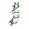
| Entry | Database: PDB / ID: 4rim | ||||||||||||||||||||
|---|---|---|---|---|---|---|---|---|---|---|---|---|---|---|---|---|---|---|---|---|---|
| Title | Native structure of intercalation-locked DNA tetraplex | ||||||||||||||||||||
 Components Components | DNA (5'-D(* Keywords KeywordsDNA / Homo-duplex / Intercalation-locked DNA tetraplex | Function / homology | DNA / DNA (> 10) |  Function and homology information Function and homology informationBiological species | synthetic construct (others) | Method |  X-RAY DIFFRACTION / SYNCHROTRON / MOLECULAR REPLACEMENT / Resolution: 2.3 Å X-RAY DIFFRACTION / SYNCHROTRON / MOLECULAR REPLACEMENT / Resolution: 2.3 Å  Authors AuthorsTripathi, S.K. / Zhang, D. / Paukstelis, P. |  CitationJournal: Nucleic Acids Res. / Year: 2015 CitationJournal: Nucleic Acids Res. / Year: 2015Title: An intercalation-locked parallel-stranded DNA tetraplex. Authors: Tripathi, S. / Zhang, D. / Paukstelis, P.J. History |
|
- Structure visualization
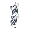
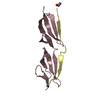
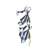
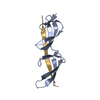
| Structure viewer | Molecule: MolmilJmol/JSmol |
|---|
- Downloads & links
-Download
| PDBx/mmCIF format | 4rim.cif.gz | 19.4 KB | Display | PDBx/mmCIF format |
|---|---|---|---|---|
| PDB format | pdb4rim.ent.gz | 12.9 KB | Display | PDB format |
| PDBx/mmJSON format | 4rim.json.gz | Tree view | PDBx/mmJSON format | |
| Others |  Other downloads Other downloads |
-Validation report
| Arichive directory | https://data.pdbj.org/pub/pdb/validation_reports/ri/4rimftp://data.pdbj.org/pub/pdb/validation_reports/ri/4rim | HTTPS FTP |
|---|
-Related structure data


-Links
 PDBj
PDBj


- Assembly
| Deposited unit | 
| ||||||||
|---|---|---|---|---|---|---|---|---|---|
| 1 | 
| ||||||||
| Unit cell |
|
-Components
| #1: DNA chain | Mass: 3373.222 Da / Num. of mol.: 1 / Source method: obtained synthetically / Source: (synth.) synthetic construct (others) |
|---|
-Experimental details
-Experiment
| Experiment | Method: X-RAY DIFFRACTION / Number of used crystals: 1 |
|---|
- Sample preparation
| Crystal | Density Matthews: 2.3 Å3/Da / Density % sol: 46.53 % |
|---|---|
| Crystal grow | Temperature: 300 K / Method: vapor diffusion, sitting drop / pH: 5.5 Details: 100 mM BaCl2, 20% MPD, 30 mM NaCaco pH 5.5, VAPOR DIFFUSION, SITTING DROP, temperature 300K |
-Data collection
| Diffraction | Mean temperature: 100 K |
|---|---|
| Diffraction source | Source: SYNCHROTRON / Site: APS  / Beamline: 24-ID-E / Wavelength: 0.9792 Å / Beamline: 24-ID-E / Wavelength: 0.9792 Å |
| Detector | Type: PSI PILATUS 6M / Detector: PIXEL / Date: Jun 30, 2014 / Details: mirror |
| Radiation | Protocol: SINGLE WAVELENGTH / Monochromatic (M) / Laue (L): M / Scattering type: x-ray |
| Radiation wavelength | Wavelength: 0.9792 Å / Relative weight: 1 |
| Reflection | Resolution: 2.3→55.94 Å / Num. obs: 1595 / % possible obs: 99 % / Observed criterion σ(F): 2 / Observed criterion σ(I): 3 / Redundancy: 7.7 % / Biso Wilson estimate: 40 Å2 / Rmerge(I) obs: 0.076 / Rsym value: 0.076 / Net I/σ(I): 2.2 |
| Reflection shell | Resolution: 2.3→2.36 Å / Redundancy: 8.2 % / Rmerge(I) obs: 0.076 / Mean I/σ(I) obs: 1.4 / Num. unique all: 378 / Rsym value: 0.487 / % possible all: 99 |
- Processing
| Software |
| ||||||||||||||||||||||||||||||||||||||||||||||||||||||||||||||||||||||||||||||||||||||||||||||||||||||||||||||||||||||||||||||||||||||||||||||||||||||||||||||||||||||||||||||||||||||
|---|---|---|---|---|---|---|---|---|---|---|---|---|---|---|---|---|---|---|---|---|---|---|---|---|---|---|---|---|---|---|---|---|---|---|---|---|---|---|---|---|---|---|---|---|---|---|---|---|---|---|---|---|---|---|---|---|---|---|---|---|---|---|---|---|---|---|---|---|---|---|---|---|---|---|---|---|---|---|---|---|---|---|---|---|---|---|---|---|---|---|---|---|---|---|---|---|---|---|---|---|---|---|---|---|---|---|---|---|---|---|---|---|---|---|---|---|---|---|---|---|---|---|---|---|---|---|---|---|---|---|---|---|---|---|---|---|---|---|---|---|---|---|---|---|---|---|---|---|---|---|---|---|---|---|---|---|---|---|---|---|---|---|---|---|---|---|---|---|---|---|---|---|---|---|---|---|---|---|---|---|---|---|---|
| Refinement | Method to determine structure: MOLECULAR REPLACEMENT / Resolution: 2.3→50 Å / Cor.coef. Fo:Fc: 0.922 / Cor.coef. Fo:Fc free: 0.902 / SU B: 13.738 / SU ML: 0.149 / Cross valid method: THROUGHOUT / ESU R: 0.358 / ESU R Free: 0.251 / Stereochemistry target values: MAXIMUM LIKELIHOOD / Details: HYDROGENS HAVE BEEN ADDED IN THE RIDING POSITIONS
| ||||||||||||||||||||||||||||||||||||||||||||||||||||||||||||||||||||||||||||||||||||||||||||||||||||||||||||||||||||||||||||||||||||||||||||||||||||||||||||||||||||||||||||||||||||||
| Solvent computation | Ion probe radii: 0.9 Å / Shrinkage radii: 0.9 Å / VDW probe radii: 1 Å / Solvent model: MASK | ||||||||||||||||||||||||||||||||||||||||||||||||||||||||||||||||||||||||||||||||||||||||||||||||||||||||||||||||||||||||||||||||||||||||||||||||||||||||||||||||||||||||||||||||||||||
| Displacement parameters | Biso mean: 59.175 Å2
| ||||||||||||||||||||||||||||||||||||||||||||||||||||||||||||||||||||||||||||||||||||||||||||||||||||||||||||||||||||||||||||||||||||||||||||||||||||||||||||||||||||||||||||||||||||||
| Refinement step | Cycle: LAST / Resolution: 2.3→50 Å
| ||||||||||||||||||||||||||||||||||||||||||||||||||||||||||||||||||||||||||||||||||||||||||||||||||||||||||||||||||||||||||||||||||||||||||||||||||||||||||||||||||||||||||||||||||||||
| Refine LS restraints |
|