 Movie
Movie Controller

[English] 日本語
 Yorodumi
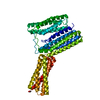
- PDB-4rhz: Crystal structure of Cry23Aa1 and Cry37Aa1 binary protein complex -
+ Open data

- Basic information
| Entry | Database: PDB / ID: 4rhz | ||||||
|---|---|---|---|---|---|---|---|
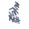
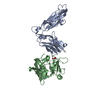
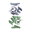
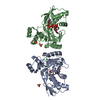
| Title | Crystal structure of Cry23Aa1 and Cry37Aa1 binary protein complex | ||||||
 Components Components |
| ||||||
 Keywords Keywords | TOXIN / aerolysin-family fold / C2-fold | ||||||
| Function / homology | Insecticidal delta endotoxin / Aerolysin-like toxin / Clostridium epsilon toxin ETX/Bacillus mosquitocidal toxin MTX2 / metal ion binding / Crystal protein / Cry23 Function and homology information Function and homology information | ||||||
| Biological species |  | ||||||
| Method |  X-RAY DIFFRACTION / SYNCHROTRON / MIR / Resolution: 2.35 Å X-RAY DIFFRACTION / SYNCHROTRON / MIR / Resolution: 2.35 Å | ||||||
 Authors Authors | Rydel, T.J. / Williams, J.M. / Brown, G.R. / Guzov, V.M. / Sturman, E.J. / Evdokimov, A. | ||||||
 Citation Citation | Journal: To be Published Title: The Associated Bacillus thuringiensis Binary Protein Complex of Cry23Aa1 and Cry37Aa1: Crystal Structure, Insecticidal Data, and Pore Formation Modeling. Authors: Rydel, T.J. / Sivasupramanian, S. / Williams, J.M. / Brown, G.R. / Fu, X. / Sturman, E.J. / Roberts, J.K. / Guzov, V.M. / Seale, J.W. / Moshiri, F.M. / Zheng, M. / Halls, C. / Evdokimov, A. | ||||||
| History |
|
- Structure visualization
| Structure viewer | Molecule: MolmilJmol/JSmol |
|---|
- Downloads & links
-Download
| PDBx/mmCIF format | 4rhz.cif.gz | 94.5 KB | Display | PDBx/mmCIF format |
|---|---|---|---|---|
| PDB format | pdb4rhz.ent.gz | 72.1 KB | Display | PDB format |
| PDBx/mmJSON format | 4rhz.json.gz | Tree view | PDBx/mmJSON format | |
| Others |  Other downloads Other downloads |
-Validation report
| Summary document | 4rhz_validation.pdf.gz | 441.1 KB | Display | wwPDB validaton report |
|---|---|---|---|---|
| Full document | 4rhz_full_validation.pdf.gz | 451.1 KB | Display | |
| Data in XML | 4rhz_validation.xml.gz | 21.5 KB | Display | |
| Data in CIF | 4rhz_validation.cif.gz | 29.3 KB | Display | |
| Arichive directory | https://data.pdbj.org/pub/pdb/validation_reports/rh/4rhzftp://data.pdbj.org/pub/pdb/validation_reports/rh/4rhz | HTTPS FTP |
-Related structure data
| Similar structure data |
|---|


-Links
 PDBj
PDBj- Assembly
| Deposited unit | 
| ||||||||
|---|---|---|---|---|---|---|---|---|---|
| 1 |
| ||||||||
| Unit cell |
|
-Components
| #1: Protein | Mass: 29235.285 Da / Num. of mol.: 1 Source method: isolated from a genetically manipulated source Source: (gene. exp.) |
|---|---|
| #2: Protein | Mass: 14191.533 Da / Num. of mol.: 1 Source method: isolated from a genetically manipulated source Source: (gene. exp.) |
| #3: Chemical | ChemComp-ZN /   Mass: 65.409 Da / Num. of mol.: 1 / Source method: obtained synthetically / Formula: Zn Mass: 65.409 Da / Num. of mol.: 1 / Source method: obtained synthetically / Formula: Zn |
| #4: Chemical | ChemComp-CA /   Mass: 40.078 Da / Num. of mol.: 1 / Source method: obtained synthetically / Formula: Ca Mass: 40.078 Da / Num. of mol.: 1 / Source method: obtained synthetically / Formula: Ca |
| #5: Water | ChemComp-HOH /  Mass: 18.015 Da / Num. of mol.: 209 / Source method: isolated from a natural source / Formula: H2O Mass: 18.015 Da / Num. of mol.: 209 / Source method: isolated from a natural source / Formula: H2O |
| Has protein modification | Y |
-Experimental details
-Experiment
| Experiment | Method: X-RAY DIFFRACTION / Number of used crystals: 1 |
|---|
- Sample preparation
| Crystal | Density Matthews: 4.21 Å3/Da / Density % sol: 70.76 % |
|---|---|
| Crystal grow | Temperature: 296 K / Method: vapor diffusion, hanging drop / pH: 8.5 Details: The protein sample was at a concentration of 2.1mg/mL in 25mM ethanolamine, and was prepared from chromatography fractions containing the co-eluted protein complex. For vapor diffusion ...Details: The protein sample was at a concentration of 2.1mg/mL in 25mM ethanolamine, and was prepared from chromatography fractions containing the co-eluted protein complex. For vapor diffusion crystallization, the precipitant solution was 2-6%(w/v) PEG8000 in 0.1M Tris-pH 8.5 buffer. A droplet using 2uL of protein and 2uL of precipitant solution was placed over a well containing 500 uL of precipitant solution. Crystals appeared within one week., VAPOR DIFFUSION, HANGING DROP, temperature 296K |
-Data collection
| Diffraction | Mean temperature: 100 K |
|---|---|
| Diffraction source | Source: SYNCHROTRON / Site: APS  / Beamline: 17-ID / Wavelength: 1 Å / Beamline: 17-ID / Wavelength: 1 Å |
| Detector | Type: MARRESEARCH / Detector: CCD / Date: Mar 15, 2000 |
| Radiation | Protocol: SINGLE WAVELENGTH / Monochromatic (M) / Laue (L): M / Scattering type: x-ray |
| Radiation wavelength | Wavelength: 1 Å / Relative weight: 1 |
| Reflection | Resolution: 2.35→109 Å / Num. all: 30246 / Num. obs: 30216 / % possible obs: 99.9 % / Redundancy: 10.1 % / Rmerge(I) obs: 0.05 / Net I/σ(I): 49.1 |
- Processing
| Software |
| ||||||||||||||||||||||||||||||||||||||||||||||||||||||||||||||||||||||||||||||||||||||||||||||||||||||||||||||||||||||||||||||||||||||||||||||||||||||||||||||||||||||||||||||||||||||
|---|---|---|---|---|---|---|---|---|---|---|---|---|---|---|---|---|---|---|---|---|---|---|---|---|---|---|---|---|---|---|---|---|---|---|---|---|---|---|---|---|---|---|---|---|---|---|---|---|---|---|---|---|---|---|---|---|---|---|---|---|---|---|---|---|---|---|---|---|---|---|---|---|---|---|---|---|---|---|---|---|---|---|---|---|---|---|---|---|---|---|---|---|---|---|---|---|---|---|---|---|---|---|---|---|---|---|---|---|---|---|---|---|---|---|---|---|---|---|---|---|---|---|---|---|---|---|---|---|---|---|---|---|---|---|---|---|---|---|---|---|---|---|---|---|---|---|---|---|---|---|---|---|---|---|---|---|---|---|---|---|---|---|---|---|---|---|---|---|---|---|---|---|---|---|---|---|---|---|---|---|---|---|---|
| Refinement | Method to determine structure: MIR / Resolution: 2.35→49.5 Å / Cor.coef. Fo:Fc: 0.951 / Cor.coef. Fo:Fc free: 0.927 / Cross valid method: THROUGHOUT / ESU R: 0.21 / ESU R Free: 0.198 / Stereochemistry target values: MAXIMUM LIKELIHOOD / Details: HYDROGENS HAVE BEEN ADDED IN THE RIDING POSITIONS
| ||||||||||||||||||||||||||||||||||||||||||||||||||||||||||||||||||||||||||||||||||||||||||||||||||||||||||||||||||||||||||||||||||||||||||||||||||||||||||||||||||||||||||||||||||||||
| Solvent computation | Ion probe radii: 0.8 Å / Shrinkage radii: 0.8 Å / VDW probe radii: 1.4 Å / Solvent model: MASK | ||||||||||||||||||||||||||||||||||||||||||||||||||||||||||||||||||||||||||||||||||||||||||||||||||||||||||||||||||||||||||||||||||||||||||||||||||||||||||||||||||||||||||||||||||||||
| Displacement parameters | Biso mean: 42.6 Å2
| ||||||||||||||||||||||||||||||||||||||||||||||||||||||||||||||||||||||||||||||||||||||||||||||||||||||||||||||||||||||||||||||||||||||||||||||||||||||||||||||||||||||||||||||||||||||
| Refinement step | Cycle: LAST / Resolution: 2.35→49.5 Å
| ||||||||||||||||||||||||||||||||||||||||||||||||||||||||||||||||||||||||||||||||||||||||||||||||||||||||||||||||||||||||||||||||||||||||||||||||||||||||||||||||||||||||||||||||||||||
| Refine LS restraints |
|