 Movie
Movie Controller

[English] 日本語
 Yorodumi
- PDB-4rd7: The crystal structure of a Cupin 2 conserved barrel domain protei... -
+ Open data

- Basic information
| Entry | Database: PDB / ID: 4rd7 | ||||||
|---|---|---|---|---|---|---|---|
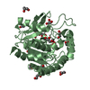
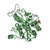
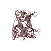
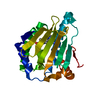
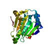
| Title | The crystal structure of a Cupin 2 conserved barrel domain protein from Salinispora arenicola CNS-205 | ||||||
 Components Components | Cupin 2 conserved barrel domain protein | ||||||
 Keywords Keywords | UNKNOWN FUNCTION / structural genomics / PSI-Biology / protein structure initiative / midwest center for structural genomics / MCSG / Enzyme Discovery for Natural Product Biosynthesis / NatPro | ||||||
| Function / homology |  Function and homology information Function and homology information: / Cupin 2, conserved barrel / Cupin domain / RmlC-like cupin domain superfamily / Jelly Rolls / RmlC-like jelly roll fold / Jelly Rolls / Sandwich / Mainly Beta Similarity search - Domain/homology | ||||||
| Biological species |  Salinispora arenicola (bacteria) Salinispora arenicola (bacteria) | ||||||
| Method |  X-RAY DIFFRACTION / SYNCHROTRON / SAD / Resolution: 1.571 Å X-RAY DIFFRACTION / SYNCHROTRON / SAD / Resolution: 1.571 Å | ||||||
 Authors Authors | Tan, K. / Gu, M. / Clancy, S. / Phillips Jr., G.N. / Joachimiak, A. / Midwest Center for Structural Genomics (MCSG) / Enzyme Discovery for Natural Product Biosynthesis (NatPro) | ||||||
 Citation Citation | Journal: To be Published Title: The crystal structure of a Cupin 2 conserved barrel domain protein from Salinispora arenicola CNS-205 Authors: Tan, K. / Gu, M. / Clancy, S. / Phillips Jr., G.N. / Joachimiak, A. | ||||||
| History |
|
- Structure visualization
| Structure viewer | Molecule: MolmilJmol/JSmol |
|---|
- Downloads & links
-Download
| PDBx/mmCIF format | 4rd7.cif.gz | 66.6 KB | Display | PDBx/mmCIF format |
|---|---|---|---|---|
| PDB format | pdb4rd7.ent.gz | 49.2 KB | Display | PDB format |
| PDBx/mmJSON format | 4rd7.json.gz | Tree view | PDBx/mmJSON format | |
| Others |  Other downloads Other downloads |
-Validation report
| Arichive directory | https://data.pdbj.org/pub/pdb/validation_reports/rd/4rd7ftp://data.pdbj.org/pub/pdb/validation_reports/rd/4rd7 | HTTPS FTP |
|---|
-Related structure data
| Similar structure data | |
|---|---|
| Other databases |

-Links
 PDBj
PDBj- Assembly
| Deposited unit | 
| ||||||||
|---|---|---|---|---|---|---|---|---|---|
| 1 | 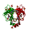
| ||||||||
| Unit cell |
|
-Components
| #1: Protein | Mass: 14571.407 Da / Num. of mol.: 1 Source method: isolated from a genetically manipulated source Source: (gene. exp.) Salinispora arenicola (bacteria) / Strain: CNS-205 / Gene: Sare_2150 / Plasmid: pMCSG68 / Production host: | ||||||
|---|---|---|---|---|---|---|---|
| #2: Chemical | ChemComp-SO4 /   Mass: 96.063 Da / Num. of mol.: 5 / Source method: obtained synthetically / Formula: SO4 Mass: 96.063 Da / Num. of mol.: 5 / Source method: obtained synthetically / Formula: SO4#3: Chemical | ChemComp-GOL / |   Mass: 92.094 Da / Num. of mol.: 1 / Source method: obtained synthetically / Formula: C3H8O3 Mass: 92.094 Da / Num. of mol.: 1 / Source method: obtained synthetically / Formula: C3H8O3#4: Water | ChemComp-HOH / |  Mass: 18.015 Da / Num. of mol.: 98 / Source method: isolated from a natural source / Formula: H2O Mass: 18.015 Da / Num. of mol.: 98 / Source method: isolated from a natural source / Formula: H2OHas protein modification | Y | |
-Experimental details
-Experiment
| Experiment | Method: X-RAY DIFFRACTION / Number of used crystals: 1 |
|---|
- Sample preparation
| Crystal | Density Matthews: 2.17 Å3/Da / Density % sol: 43.43 % |
|---|---|
| Crystal grow | Temperature: 289 K / Method: vapor diffusion, sitting drop / pH: 4.5 Details: 1.6M Ammonium Sulfate, 0.1M MES:NaOH, 10% (v/v) Dioxane, pH 4.5, VAPOR DIFFUSION, SITTING DROP, temperature 289K |
-Data collection
| Diffraction | Mean temperature: 100 K |
|---|---|
| Diffraction source | Source: SYNCHROTRON / Site: APS  / Beamline: 19-ID / Wavelength: 0.97915 Å / Beamline: 19-ID / Wavelength: 0.97915 Å |
| Detector | Type: ADSC QUANTUM 315r / Detector: CCD / Date: Aug 25, 2014 / Details: mirror |
| Radiation | Monochromator: Si 111 crystal / Protocol: SINGLE WAVELENGTH / Monochromatic (M) / Laue (L): M / Scattering type: x-ray |
| Radiation wavelength | Wavelength: 0.97915 Å / Relative weight: 1 |
| Reflection | Resolution: 1.57→36.5 Å / Num. all: 18841 / Num. obs: 18841 / % possible obs: 100 % / Observed criterion σ(F): 0 / Observed criterion σ(I): -5 / Redundancy: 14 % / Biso Wilson estimate: 13.76 Å2 / Rmerge(I) obs: 0.124 / Net I/σ(I): 47.2 |
| Reflection shell | Resolution: 1.57→1.6 Å / Redundancy: 7.9 % / Rmerge(I) obs: 0.572 / Mean I/σ(I) obs: 3.9 / Num. unique all: 891 / % possible all: 99.8 |
- Processing
| Software |
| |||||||||||||||||||||||||||||||||||||||||||||||||
|---|---|---|---|---|---|---|---|---|---|---|---|---|---|---|---|---|---|---|---|---|---|---|---|---|---|---|---|---|---|---|---|---|---|---|---|---|---|---|---|---|---|---|---|---|---|---|---|---|---|---|
| Refinement | Method to determine structure: SAD / Resolution: 1.571→36.497 Å / SU ML: 0.12 / σ(F): 1.36 / Phase error: 18.81 / Stereochemistry target values: ML
| |||||||||||||||||||||||||||||||||||||||||||||||||
| Solvent computation | Shrinkage radii: 0.9 Å / VDW probe radii: 1.11 Å / Solvent model: FLAT BULK SOLVENT MODEL | |||||||||||||||||||||||||||||||||||||||||||||||||
| Refinement step | Cycle: LAST / Resolution: 1.571→36.497 Å
| |||||||||||||||||||||||||||||||||||||||||||||||||
| Refine LS restraints |
| |||||||||||||||||||||||||||||||||||||||||||||||||
| LS refinement shell |
|
