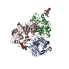
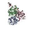
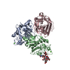
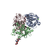
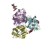
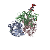
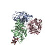
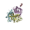
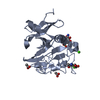
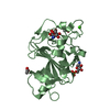
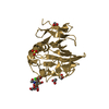
[English] 日本語
Yorodumi
- PDB-4r9j: L-ficolin complexed to glucosamine-6-sulfate -

+
Open data


ID or keywords:

Loading...

-
Basic information

Entry
Database: PDB / ID: 4r9j
TitleL-ficolin complexed to glucosamine-6-sulfate
ComponentsFicolin-2
KeywordsSUGAR BINDING PROTEIN / FIBRINOGEN-LIKE domain / INNATE IMMUNITY / PATTERN RECOGNITION PROTEIN / LECTIN / IMMUNOLOGY / LECTIN-LIKE / plasma / extracellular
Function / homology
Function and homology information


mannan binding / recognition of apoptotic cell / Ficolins bind to repetitive carbohydrate structures on the target cell surface / Lectin pathway of complement activation / positive regulation of opsonization / cell surface pattern recognition receptor signaling pathway / opsonization / complement activation, lectin pathway / carbohydrate derivative binding / collagen trimer ...mannan binding / recognition of apoptotic cell / Ficolins bind to repetitive carbohydrate structures on the target cell surface / Lectin pathway of complement activation / positive regulation of opsonization / cell surface pattern recognition receptor signaling pathway / opsonization / complement activation, lectin pathway / carbohydrate derivative binding / collagen trimer / serine-type endopeptidase complex / proteoglycan binding / Initial triggering of complement / antigen binding / calcium-dependent protein binding / collagen-containing extracellular matrix / defense response to Gram-negative bacterium / blood microparticle / defense response to Gram-positive bacterium / external side of plasma membrane / signaling receptor binding / proteolysis / extracellular space / extracellular exosome / extracellular region / metal ion binding
Similarity search - Function
: / Gamma-fibrinogen Carboxyl Terminal Fragment; domain 2 / Gamma-fibrinogen Carboxyl Terminal Fragment, domain 2 / Gamma Fibrinogen; Chain A, domain 1 / Gamma Fibrinogen, chain A, domain 1 / Fibrinogen, conserved site / Fibrinogen C-terminal domain signature. / Fibrinogen-related domains (FReDs) / Fibrinogen, alpha/beta/gamma chain, C-terminal globular, subdomain 1 / Fibrinogen beta and gamma chains, C-terminal globular domain ...: / Gamma-fibrinogen Carboxyl Terminal Fragment; domain 2 / Gamma-fibrinogen Carboxyl Terminal Fragment, domain 2 / Gamma Fibrinogen; Chain A, domain 1 / Gamma Fibrinogen, chain A, domain 1 / Fibrinogen, conserved site / Fibrinogen C-terminal domain signature. / Fibrinogen-related domains (FReDs) / Fibrinogen, alpha/beta/gamma chain, C-terminal globular, subdomain 1 / Fibrinogen beta and gamma chains, C-terminal globular domain / Fibrinogen, alpha/beta/gamma chain, C-terminal globular domain / Fibrinogen-like, C-terminal / Fibrinogen C-terminal domain profile. / Collagen triple helix repeat / Collagen triple helix repeat (20 copies) / Few Secondary Structures / Irregular / Alpha-Beta Complex / Alpha Beta
Similarity search - Domain/homology
2-amino-2-deoxy-6-O-sulfo-alpha-D-glucopyranose / ACETATE ION / Ficolin-2
Similarity search - Component
Biological speciesHomo sapiens (human)
MethodX-RAY DIFFRACTION / SYNCHROTRON / MOLECULAR REPLACEMENT / Resolution: 2.1 Å
AuthorsLaffly, E. / Lacroix, M. / Martin, L. / Vassal-Stermann, E. / Thielens, N. / Gaboriaud, C.
CitationJournal: Febs Lett. / Year: 2014
Title: Human ficolin-2 recognition versatility extended: An update on the binding of ficolin-2 to sulfated/phosphated carbohydrates.
Authors: Laffly, E. / Lacroix, M. / Martin, L. / Vassal-Stermann, E. / Thielens, N.M. / Gaboriaud, C.
History
DepositionSep 5, 2014Deposition site: RCSB / Processing site: RCSB
Revision 1.0Nov 5, 2014Provider: repository / Type: Initial release
Revision 1.1Dec 17, 2014Group: Database references
Revision 1.2Jan 14, 2015Group: Database references
Revision 2.0Jul 29, 2020Group: Atomic model / Data collection ...Atomic model / Data collection / Derived calculations / Structure summary
Category: atom_site / atom_site_anisotrop ...atom_site / atom_site_anisotrop / chem_comp / entity / pdbx_branch_scheme / pdbx_chem_comp_identifier / pdbx_entity_branch / pdbx_entity_branch_descriptor / pdbx_entity_branch_link / pdbx_entity_branch_list / pdbx_entity_nonpoly / pdbx_nonpoly_scheme / pdbx_struct_assembly_gen / pdbx_struct_conn_angle / pdbx_struct_special_symmetry / struct_asym / struct_conn / struct_site / struct_site_gen
Item: _atom_site.B_iso_or_equiv / _atom_site.Cartn_x ..._atom_site.B_iso_or_equiv / _atom_site.Cartn_x / _atom_site.Cartn_y / _atom_site.Cartn_z / _atom_site.auth_asym_id / _atom_site.auth_atom_id / _atom_site.auth_comp_id / _atom_site.auth_seq_id / _atom_site.label_asym_id / _atom_site.label_atom_id / _atom_site.label_comp_id / _atom_site.label_entity_id / _atom_site.type_symbol / _atom_site_anisotrop.U[1][1] / _atom_site_anisotrop.U[1][2] / _atom_site_anisotrop.U[1][3] / _atom_site_anisotrop.U[2][2] / _atom_site_anisotrop.U[2][3] / _atom_site_anisotrop.U[3][3] / _atom_site_anisotrop.pdbx_auth_asym_id / _atom_site_anisotrop.pdbx_auth_atom_id / _atom_site_anisotrop.pdbx_auth_comp_id / _atom_site_anisotrop.pdbx_auth_seq_id / _atom_site_anisotrop.pdbx_label_asym_id / _atom_site_anisotrop.pdbx_label_atom_id / _atom_site_anisotrop.pdbx_label_comp_id / _atom_site_anisotrop.type_symbol / _chem_comp.name / _chem_comp.type / _entity.formula_weight / _entity.pdbx_description / _entity.pdbx_number_of_molecules / _entity.src_method / _entity.type / _pdbx_struct_assembly_gen.asym_id_list / _pdbx_struct_conn_angle.ptnr1_auth_asym_id / _pdbx_struct_conn_angle.ptnr1_auth_comp_id / _pdbx_struct_conn_angle.ptnr1_auth_seq_id / _pdbx_struct_conn_angle.ptnr1_label_asym_id / _pdbx_struct_conn_angle.ptnr1_label_atom_id / _pdbx_struct_conn_angle.ptnr1_label_comp_id / _pdbx_struct_conn_angle.ptnr1_label_seq_id / _pdbx_struct_conn_angle.ptnr2_auth_asym_id / _pdbx_struct_conn_angle.ptnr2_auth_seq_id / _pdbx_struct_conn_angle.ptnr2_label_asym_id / _pdbx_struct_conn_angle.ptnr3_auth_asym_id / _pdbx_struct_conn_angle.ptnr3_auth_comp_id / _pdbx_struct_conn_angle.ptnr3_auth_seq_id / _pdbx_struct_conn_angle.ptnr3_label_asym_id / _pdbx_struct_conn_angle.ptnr3_label_atom_id / _pdbx_struct_conn_angle.ptnr3_label_comp_id / _pdbx_struct_conn_angle.ptnr3_label_seq_id / _pdbx_struct_conn_angle.value / _pdbx_struct_special_symmetry.label_asym_id / _struct_conn.pdbx_dist_value / _struct_conn.pdbx_leaving_atom_flag / _struct_conn.pdbx_role / _struct_conn.ptnr1_auth_asym_id / _struct_conn.ptnr1_auth_comp_id / _struct_conn.ptnr1_auth_seq_id / _struct_conn.ptnr1_label_asym_id / _struct_conn.ptnr1_label_atom_id / _struct_conn.ptnr1_label_comp_id / _struct_conn.ptnr1_label_seq_id / _struct_conn.ptnr2_auth_asym_id / _struct_conn.ptnr2_auth_comp_id / _struct_conn.ptnr2_auth_seq_id / _struct_conn.ptnr2_label_asym_id / _struct_conn.ptnr2_label_atom_id / _struct_conn.ptnr2_label_comp_id
Description: Carbohydrate remediation / Provider: repository / Type: Remediation

-
Structure visualization

Structure viewerMolecule:
MolmilJmol/JSmol

Downloads & links

-
Assembly

Deposited unit
G: Ficolin-2
A: Ficolin-2
B: Ficolin-2
hetero molecules


Theoretical massNumber of molelcules
Total (without water)76,08920
Polymers73,8343
Non-polymers2,25517
Water4,216234
1


  • Idetical with deposited unit
  • defined by author
TypeNameSymmetry operationNumber
identity operation1_555x,y,z1
2
A: Ficolin-2
hetero molecules


Theoretical massNumber of molelcules
Total (without water)25,2907
Polymers24,6111
Non-polymers6796
Water181
TypeNameSymmetry operationNumber
identity operation1_555x,y,z1
MethodPISA
3
B: Ficolin-2
hetero molecules


Theoretical massNumber of molelcules
Total (without water)25,2886
Polymers24,6111
Non-polymers6775
Water181
TypeNameSymmetry operationNumber
identity operation1_555x,y,z1
MethodPISA
4
G: Ficolin-2
hetero molecules


Theoretical massNumber of molelcules
Total (without water)25,5117
Polymers24,6111
Non-polymers9006
Water181
TypeNameSymmetry operationNumber
identity operation1_555x,y,z1
MethodPISA
Unit cell
Length a, b, c (Å)97.170, 97.170, 139.990
Angle α, β, γ (deg.)90.00, 90.00, 120.00
Int Tables number154
Space group name H-MP3221
Components on special symmetry positions
IDModelComponents
11G-486-

HOH

Noncrystallographic symmetry (NCS)NCS domain:
IDEns-IDDetails
11G
21A
12G
22B
13A
23B

NCS domain segments:
Dom-IDComponent-IDEns-IDRefine codeAuth asym-IDAuth seq-ID
1010G72 - 288
2010A72 - 288
1020G72 - 288
2020B72 - 288
1030A72 - 288
2030B72 - 288

NCS ensembles :
ID
1
2
3

-
Components

-
Protein , 1 types, 3 molecules GAB

#1: Protein Ficolin-2 / 37 kDa elastin-binding protein / Collagen/fibrinogen domain-containing protein 2 / EBP-37 / Ficolin- ...37 kDa elastin-binding protein / Collagen/fibrinogen domain-containing protein 2 / EBP-37 / Ficolin-B / Ficolin-beta / Hucolin / L-ficolin / Serum lectin p35


Mass: 24611.273 Da / Num. of mol.: 3 / Fragment: SUGAR BINDING DOMAIN (UNP RESIDUES 97-313)
Source method: isolated from a genetically manipulated source
Source: (gene. exp.) Homo sapiens (human) / Gene: FCN2, FCNL / Production host: 
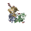
Unidentified Baculovirus / References: UniProt: Q15485

-
Sugars , 3 types, 5 molecules

#2: Polysaccharide beta-D-mannopyranose-(1-4)-2-acetamido-2-deoxy-beta-D-glucopyranose-(1-4)-2-acetamido-2-deoxy-beta- ...beta-D-mannopyranose-(1-4)-2-acetamido-2-deoxy-beta-D-glucopyranose-(1-4)-2-acetamido-2-deoxy-beta-D-glucopyranose


Type: oligosaccharide / Mass: 586.542 Da / Num. of mol.: 1
Source method: isolated from a genetically manipulated source
DescriptorTypeProgram
DManpb1-4DGlcpNAcb1-4DGlcpNAcb1-Glycam Condensed SequenceGMML 1.0
WURCS=2.0/2,3,2/[a2122h-1b_1-5_2*NCC/3=O][a1122h-1b_1-5]/1-1-2/a4-b1_b4-c1WURCSPDB2Glycan 1.1.0
[]{[(4+1)][b-D-GlcpNAc]{[(4+1)][b-D-GlcpNAc]{[(4+1)][b-D-Manp]{}}}}LINUCSPDB-CARE
#6: Sugar ChemComp-3LJ / 2-amino-2-deoxy-6-O-sulfo-alpha-D-glucopyranose


Type: D-saccharide, alpha linking / Mass: 259.234 Da / Num. of mol.: 3 / Source method: obtained synthetically / Formula: C6H13NO8S
IdentifierTypeProgram
DGlcpN[6S]aCONDENSED IUPAC CARBOHYDRATE SYMBOLGMML 1.0
6-sulfo-a-D-glucopyranose-osamineCOMMON NAMEGMML 1.0
a-D-GlcpN6SO3IUPAC CARBOHYDRATE SYMBOLPDB-CARE 1.0
#7: Sugar ChemComp-NAG / 2-acetamido-2-deoxy-beta-D-glucopyranose


Type: D-saccharide, beta linking / Mass: 221.208 Da / Num. of mol.: 1
Source method: isolated from a genetically manipulated source
Formula: C8H15NO6
IdentifierTypeProgram
DGlcpNAcbCONDENSED IUPAC CARBOHYDRATE SYMBOLGMML 1.0
N-acetyl-b-D-glucopyranosamineCOMMON NAMEGMML 1.0
b-D-GlcpNAcIUPAC CARBOHYDRATE SYMBOLPDB-CARE 1.0
GlcNAcSNFG CARBOHYDRATE SYMBOLGMML 1.0

-
Non-polymers , 4 types, 246 molecules

#3: Chemical
ChemComp-CA / CALCIUM ION


Mass: 40.078 Da / Num. of mol.: 4 / Source method: obtained synthetically / Formula: Ca
#4: Chemical
ChemComp-ACT / ACETATE ION


Mass: 59.044 Da / Num. of mol.: 7 / Source method: obtained synthetically / Formula: C2H3O2
#5: Chemical ChemComp-SO4 / SULFATE ION


Mass: 96.063 Da / Num. of mol.: 1 / Source method: obtained synthetically / Formula: SO4
#8: Water ChemComp-HOH / water


Mass: 18.015 Da / Num. of mol.: 234 / Source method: isolated from a natural source / Formula: H2O

-
Experimental details

-
Experiment

ExperimentMethod: X-RAY DIFFRACTION / Number of used crystals: 1

-
Sample preparation

CrystalDensity Matthews: 2.58 Å3/Da / Density % sol: 52.4 %
Crystal growTemperature: 293 K / Method: evaporation / pH: 7
Details: Protien solution: 5.7 mg/ml in 145 mM NaCl, 50 mM triethanolamine-HCl, pH 7.4. Reservoir solution composed of 15% (w/v) PEG 8000, 200 mM Ca acetate and 0.1 M HEPES. Cryoprotection was ...Details: Protien solution: 5.7 mg/ml in 145 mM NaCl, 50 mM triethanolamine-HCl, pH 7.4. Reservoir solution composed of 15% (w/v) PEG 8000, 200 mM Ca acetate and 0.1 M HEPES. Cryoprotection was achieved by adding PEG 400 to the drop just before flash-cooling the crystal in liquid nitrogen, EVAPORATION, temperature 293K

-
Data collection

DiffractionMean temperature: 100 K
Diffraction sourceSource: SYNCHROTRON / Site: ESRF / Beamline: ID23-2 / Wavelength: 0.8726 Å
DetectorType: MARMOSAIC 225 mm CCD / Detector: CCD / Date: Mar 11, 2011
RadiationMonochromator: Si 111 / Protocol: SINGLE WAVELENGTH / Monochromatic (M) / Laue (L): M / Scattering type: x-ray
Radiation wavelengthWavelength: 0.8726 Å / Relative weight: 1
ReflectionResolution: 2.1→20 Å / Num. all: 45239 / Num. obs: 45165 / % possible obs: 99.83 % / Observed criterion σ(F): 0 / Observed criterion σ(I): -3
Reflection shell
Resolution (Å)Diffraction-ID% possible all
2.1-2.151100
2.15-2.211100
2.21-2.281100
2.28-2.351100
2.35-2.421100
2.42-2.511100
2.51-2.61100
2.6-2.711100
2.71-2.831100
2.83-2.971100
2.97-3.131100
3.13-3.321100
3.32-3.551100
3.55-3.831100
3.83-4.2199.9
4.2-4.71100
4.7-5.421100
5.42-6.641100
6.64-9.41100
9.4-20187.5

-
Processing

Software
NameVersionClassification
MAR345dtbdata collection
PHASERphasing
REFMAC5.7.0032refinement
XDSdata reduction
XSCALEdata scaling
RefinementMethod to determine structure: MOLECULAR REPLACEMENT / Resolution: 2.1→19.96 Å / Cor.coef. Fo:Fc: 0.949 / Cor.coef. Fo:Fc free: 0.931 / SU B: 7.154 / SU ML: 0.098 / Cross valid method: THROUGHOUT / ESU R: 0.194 / ESU R Free: 0.158 / Stereochemistry target values: MAXIMUM LIKELIHOOD / Details: HYDROGENS HAVE BEEN ADDED IN THE RIDING POSITIONS
RfactorNum. reflection% reflectionSelection details
Rfree0.20336 2259 5 %RANDOM
Rwork0.17238 ---
obs0.17394 42907 99.83 %-
all-42907 --
Solvent computationIon probe radii: 0.8 Å / Shrinkage radii: 0.8 Å / VDW probe radii: 1.2 Å / Solvent model: MASK
Displacement parametersBiso mean: 21.116 Å2
Baniso -1Baniso -2Baniso -3
1--0.02 Å2-0.02 Å20 Å2
2---0.02 Å20 Å2
3---0.06 Å2
Refinement stepCycle: LAST / Resolution: 2.1→19.96 Å
ProteinNucleic acidLigandSolventTotal
Num. atoms5205 0 138 234 5577
Refine LS restraints
Refine-IDTypeDev idealDev ideal targetNumber
X-RAY DIFFRACTIONr_bond_refined_d0.0190.0195598
X-RAY DIFFRACTIONr_bond_other_d0.0090.024922
X-RAY DIFFRACTIONr_angle_refined_deg1.9471.9357589
X-RAY DIFFRACTIONr_angle_other_deg1.5353.00311243
X-RAY DIFFRACTIONr_dihedral_angle_1_deg7.055666
X-RAY DIFFRACTIONr_dihedral_angle_2_deg29.59623.686293
X-RAY DIFFRACTIONr_dihedral_angle_3_deg14.47215842
X-RAY DIFFRACTIONr_dihedral_angle_4_deg18.561537
X-RAY DIFFRACTIONr_chiral_restr0.2010.2772
X-RAY DIFFRACTIONr_gen_planes_refined0.0120.026491
X-RAY DIFFRACTIONr_gen_planes_other0.0080.021468
X-RAY DIFFRACTIONr_nbd_refined
X-RAY DIFFRACTIONr_nbd_other
X-RAY DIFFRACTIONr_nbtor_refined
X-RAY DIFFRACTIONr_nbtor_other
X-RAY DIFFRACTIONr_xyhbond_nbd_refined
X-RAY DIFFRACTIONr_xyhbond_nbd_other
X-RAY DIFFRACTIONr_metal_ion_refined
X-RAY DIFFRACTIONr_metal_ion_other
X-RAY DIFFRACTIONr_symmetry_vdw_refined
X-RAY DIFFRACTIONr_symmetry_vdw_other
X-RAY DIFFRACTIONr_symmetry_hbond_refined
X-RAY DIFFRACTIONr_symmetry_hbond_other
X-RAY DIFFRACTIONr_symmetry_metal_ion_refined
X-RAY DIFFRACTIONr_symmetry_metal_ion_other
X-RAY DIFFRACTIONr_mcbond_it1.7561.5872669
X-RAY DIFFRACTIONr_mcbond_other1.7461.5852664
X-RAY DIFFRACTIONr_mcangle_it2.6712.3583318
X-RAY DIFFRACTIONr_mcangle_other3.0592.8523319
X-RAY DIFFRACTIONr_scbond_it2.2821.8452929
X-RAY DIFFRACTIONr_scbond_other2.6012.1892920
X-RAY DIFFRACTIONr_scangle_it
X-RAY DIFFRACTIONr_scangle_other3.9563.2024250
X-RAY DIFFRACTIONr_long_range_B_refined5.10415.5476338
X-RAY DIFFRACTIONr_long_range_B_other5.08515.5056264
X-RAY DIFFRACTIONr_rigid_bond_restr
X-RAY DIFFRACTIONr_sphericity_free
X-RAY DIFFRACTIONr_sphericity_bonded
Refine LS restraints NCS

Refine-ID: X-RAY DIFFRACTION / Type: interatomic distance / Weight position: 0.05

Ens-IDDom-IDAuth asym-IDNumberRms dev position (Å)
11G123080.12
12A123080.12
21G124290.11
22B124290.11
31A127100.1
32B127100.1
LS refinement shellResolution: 2.1→2.154 Å / Total num. of bins used: 20
RfactorNum. reflection% reflection
Rfree0.263 164 -
Rwork0.187 3119 -
obs--99.94 %
Refinement TLS params.

Method: refined / Refine-ID: X-RAY DIFFRACTION

IDL112)L122)L132)L222)L232)L332)S11 (Å °)S12 (Å °)S13 (Å °)S21 (Å °)S22 (Å °)S23 (Å °)S31 (Å °)S32 (Å °)S33 (Å °)T112)T122)T132)T222)T232)T332)Origin x (Å)Origin y (Å)Origin z (Å)
10.2240.109-0.10430.3607-0.16250.3819-0.0004-0.02640.02370.06060.02490.0741-0.0209-0.0466-0.02450.03530.02060.00130.02980.00090.028656.72-10.985-9.141
21.37810.0451-0.01160.3384-0.1560.3187-0.0144-0.0879-0.20370.01050.043-0.01280.02330.0383-0.02860.02470.01880.00360.03030.00680.036146.948-16.534-5.933
30.1901-0.2253-0.00160.60260.01210.23730.03230.02110.0487-0.00950.013-0.0268-0.07170.0305-0.04530.04160.00960.02790.03220.01570.033956.986-21.605-5.646
Refinement TLS group
IDRefine-IDRefine TLS-IDAuth asym-IDAuth seq-ID
1X-RAY DIFFRACTION1A72 - 288
2X-RAY DIFFRACTION1A301 - 306
3X-RAY DIFFRACTION2B72 - 288
4X-RAY DIFFRACTION2B301 - 305
5X-RAY DIFFRACTION3G72 - 288
6X-RAY DIFFRACTION3G301 - 308

+
About Yorodumi

-
News

-
Feb 9, 2022. New format data for meta-information of EMDB entries

New format data for meta-information of EMDB entries

  • Version 3 of the EMDB header file is now the official format.
  • The previous official version 1.9 will be removed from the archive.

Related info.:EMDB header

External links:wwPDB to switch to version 3 of the EMDB data model

-
Aug 12, 2020. Covid-19 info

Covid-19 info

URL: https://pdbjlvh1.pdbj.org/emnavi/covid19.php

New page: Covid-19 featured information page in EM Navigator.

Related info.:Covid-19 info / Mar 5, 2020. Novel coronavirus structure data

+
Mar 5, 2020. Novel coronavirus structure data

Novel coronavirus structure data

Related info.:Yorodumi Speices / Aug 12, 2020. Covid-19 info

External links:COVID-19 featured content - PDBj / Molecule of the Month (242):Coronavirus Proteases

+
Jan 31, 2019. EMDB accession codes are about to change! (news from PDBe EMDB page)

EMDB accession codes are about to change! (news from PDBe EMDB page)

  • The allocation of 4 digits for EMDB accession codes will soon come to an end. Whilst these codes will remain in use, new EMDB accession codes will include an additional digit and will expand incrementally as the available range of codes is exhausted. The current 4-digit format prefixed with “EMD-” (i.e. EMD-XXXX) will advance to a 5-digit format (i.e. EMD-XXXXX), and so on. It is currently estimated that the 4-digit codes will be depleted around Spring 2019, at which point the 5-digit format will come into force.
  • The EM Navigator/Yorodumi systems omit the EMD- prefix.

Related info.:Q: What is EMD? / ID/Accession-code notation in Yorodumi/EM Navigator

External links:EMDB Accession Codes are Changing Soon! / Contact to PDBj

+
Jul 12, 2017. Major update of PDB

Major update of PDB

  • wwPDB released updated PDB data conforming to the new PDBx/mmCIF dictionary.
  • This is a major update changing the version number from 4 to 5, and with Remediation, in which all the entries are updated.
  • In this update, many items about electron microscopy experimental information are reorganized (e.g. em_software).
  • Now, EM Navigator and Yorodumi are based on the updated data.

External links:wwPDB Remediation / Enriched Model Files Conforming to OneDep Data Standards Now Available in the PDB FTP Archive

-
Yorodumi

Thousand views of thousand structures

  • Yorodumi is a browser for structure data from EMDB, PDB, SASBDB, etc.
  • This page is also the successor to EM Navigator detail page, and also detail information page/front-end page for Omokage search.
  • The word "yorodu" (or yorozu) is an old Japanese word meaning "ten thousand". "mi" (miru) is to see.

Related info.:EMDB / PDB / SASBDB / Comparison of 3 databanks / Yorodumi Search / Aug 31, 2016. New EM Navigator & Yorodumi / Yorodumi Papers / Jmol/JSmol / Function and homology information / Changes in new EM Navigator and Yorodumi

Read more