- PDB-4r98: Chimera of the N-terminal domain of E. coli FeoB -
+
Open data
ID or keywords:
Loading...
-
Basic information
Entry
Database: PDB / ID: 4r98
Title
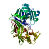
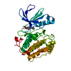
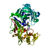
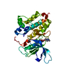
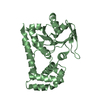
Chimera of the N-terminal domain of E. coli FeoB
Components
Ferrous iron transport protein B
Keywords
METAL TRANSPORT / FeoB
Function / homology
Function and homology information
iron ion import across plasma membrane / Metal ion assimilation from the host / ferrous iron transmembrane transporter activity / DNA damage response / GTP binding / identical protein binding / plasma membrane Similarity search - Function
Helix Hairpins - #1770 / Ferrous iron transport protein B, C-terminal / Ferrous iron transport protein B C terminus / FeoB, cytosolic helical domain / FeoB cytosolic helical domain / Ferrous iron transport protein B / FeoB-type guanine nucleotide-binding (G) domain / : / Ferrous iron transport protein B / FeoB-type guanine nucleotide-binding (G) domain profile. ...Helix Hairpins - #1770 / Ferrous iron transport protein B, C-terminal / Ferrous iron transport protein B C terminus / FeoB, cytosolic helical domain / FeoB cytosolic helical domain / Ferrous iron transport protein B / FeoB-type guanine nucleotide-binding (G) domain / : / Ferrous iron transport protein B / FeoB-type guanine nucleotide-binding (G) domain profile. / Nucleoside transporter/FeoB GTPase, Gate domain / Nucleoside recognition / Helix Hairpins / P-loop containing nucleotide triphosphate hydrolases / Rossmann fold / P-loop containing nucleoside triphosphate hydrolase / Orthogonal Bundle / 3-Layer(aba) Sandwich / Mainly Alpha / Alpha Beta Similarity search - Domain/homology
Mass: 18.015 Da / Num. of mol.: 27 / Source method: isolated from a natural source / Formula: H2O
Sequence details
THIS STRUCTURE IS REPRESENTING A MODIFIED SUB CLONE OF FEOB, WHERE A SHORT SEGMENT HAS BEEN ...THIS STRUCTURE IS REPRESENTING A MODIFIED SUB CLONE OF FEOB, WHERE A SHORT SEGMENT HAS BEEN REPLACED FOR FUNCTIONAL STUDIES
-
Experimental details
-
Experiment
Experiment
Method: X-RAY DIFFRACTION
-
Sample preparation
Crystal
Density Matthews: 2.29 Å3/Da / Density % sol: 46.32 %
Crystal grow
Method: vapor diffusion, hanging drop / pH: 6.5 Details: 22 % PEG 3350, 0.1 M Bis Tris Propane pH 6.5 and 0.2 M Sodium formate, VAPOR DIFFUSION, HANGING DROP
-
Data collection
Diffraction
ID
Crystal-ID
1
1
2
1
Diffraction source
Source
Site
Beamline
Type
ID
Wavelength (Å)
ROTATING ANODE
APS
23-ID-B
RIGAKU RU200
1
1.54
Australian Synchrotron
MX2
2
Detector
Type: MAR scanner 345 mm plate / Detector: IMAGE PLATE / Date: Sep 1, 2010
Radiation
Protocol: SINGLE WAVELENGTH / Monochromatic (M) / Laue (L): M / Scattering type: x-ray
 Movie
Movie Controller


 Open data
 Basic information
 Components
 Keywords
 Function and homology information
 X-RAY DIFFRACTION /
  Authors
 Citation
 Structure visualization
 Downloads & links
 Other downloads


 PDBj
PDBj Assembly
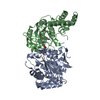

 Mass: 442.216 Da / Num. of mol.: 1 / Source method: obtained synthetically / Formula: C10H16N6O10P2
 Mass: 18.015 Da / Num. of mol.: 27 / Source method: isolated from a natural source / Formula: H2O
 Sample preparation

 Processing