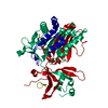
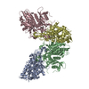
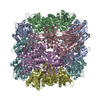
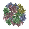
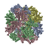
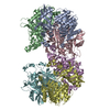
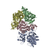
ジャーナル: Autophagy / 年: 2015 タイトル: Structure of yeast Ape1 and its role in autophagic vesicle formation. 著者: Ming-Yuan Su / Wen-Hsin Peng / Meng-Ru Ho / Shih-Chieh Su / Yuan-Chih Chang / Guang-Chao Chen / Chung-I Chang / 要旨: In Saccharomyces cerevisiae, a constitutive biosynthetic transport pathway, termed the cytoplasm-to-vacuole targeting (Cvt) pathway, sequesters precursor aminopeptidase I (prApe1) dodecamers in the ...In Saccharomyces cerevisiae, a constitutive biosynthetic transport pathway, termed the cytoplasm-to-vacuole targeting (Cvt) pathway, sequesters precursor aminopeptidase I (prApe1) dodecamers in the form of a large complex into a Cvt vesicle using autophagic machinery, targeting it into the vacuole (the yeast lysosome) where it is proteolytically processed into its mature form, Ape1, by removal of an amino-terminal 45-amino acid propeptide. prApe1 is thought to serve as a scaffolding cargo critical for the assembly of the Cvt vesicle by presenting the propeptide to mediate higher-ordered complex formation and autophagic receptor recognition. Here we report the X-ray crystal structure of Ape1 at 2.5 Å resolution and reveal its dodecameric architecture consisting of dimeric and trimeric units, which associate to form a large tetrahedron. The propeptide of prApe1 exhibits concentration-dependent oligomerization and forms a stable tetramer. Structure-based mutagenesis demonstrates that disruption of the inter-subunit interface prevents dodecameric assembly and vacuolar targeting in vivo despite the presence of the propeptide. Furthermore, by examining the vacuolar import of propeptide-fused exogenous protein assemblies with different quaternary structures, we found that 3-dimensional spatial distribution of propeptides presented by a scaffolding cargo is essential for the assembly of the Cvt vesicle for vacuolar delivery. This study describes a molecular framework for understanding the mechanism of Cvt or autophagosomal biogenesis in selective macroautophagy.
解像度: 2.5→116.23 Å / Cor.coef. Fo:Fc: 0.927 / Cor.coef. Fo:Fc free: 0.9 / SU B: 9.891 / SU ML: 0.216 / 交差検証法: THROUGHOUT / ESU R: 0.402 / ESU R Free: 0.261 / 立体化学のターゲット値: MAXIMUM LIKELIHOOD / 詳細: HYDROGENS HAVE BEEN ADDED IN THE RIDING POSITIONS
Rfactor
反射数
%反射
Selection details
Rfree
0.24637
4222
5 %
RANDOM
Rwork
0.21532
-
-
-
obs
0.2169
80667
95.69 %
-
溶媒の処理
イオンプローブ半径: 0.8 Å / 減衰半径: 0.8 Å / VDWプローブ半径: 1.2 Å / 溶媒モデル: MASK
 ムービー
ムービー コントローラー


 データを開く
 基本情報
基本情報 要素
要素 キーワード
キーワード 機能・相同性情報
 X線回折 /
  データ登録者
 引用
引用
 構造の表示
構造の表示 ダウンロードとリンク
 その他のダウンロード
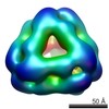

 PDBj
PDBj
 集合体
集合体

 分子量: 18.015 Da / 分子数: 91 / 由来タイプ: 天然 / 式: H2O
 試料調製
試料調製 解析
解析