 Movie
Movie Controller


+ Open data

- Basic information
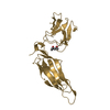
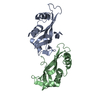
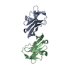
| Entry | Database: PDB / ID: 4r62 | ||||||
|---|---|---|---|---|---|---|---|
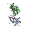
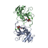
| Title | Structure of Rad6~Ub | ||||||
 Components Components |
| ||||||
 Keywords Keywords | NUCLEAR PROTEIN / E2 conjugating enzyme / UBC / Monoubiquitination of Histone H2B at K123 in Saccharomyces cerevisiae / Bre1 / Ubiquitin / PCNA / Rad18 / Histone H2B / UBIQUITINATION / NUCLEUS | ||||||
| Function / homology |  Function and homology information Function and homology informationMUB1-RAD6-UBR2 ubiquitin ligase complex / RAD6-UBR2 ubiquitin ligase complex / Rad6-Rad18 complex / regulation of dipeptide transport / UBR1-RAD6 ubiquitin ligase complex / sno(s)RNA transcription / HULC complex / error-free postreplication DNA repair / stress-induced homeostatically regulated protein degradation pathway / : ...MUB1-RAD6-UBR2 ubiquitin ligase complex / RAD6-UBR2 ubiquitin ligase complex / Rad6-Rad18 complex / regulation of dipeptide transport / UBR1-RAD6 ubiquitin ligase complex / sno(s)RNA transcription / HULC complex / error-free postreplication DNA repair / stress-induced homeostatically regulated protein degradation pathway / : / meiotic DNA double-strand break formation / ubiquitin-dependent protein catabolic process via the N-end rule pathway / cytoplasm protein quality control by the ubiquitin-proteasome system / : / telomere maintenance via recombination / error-free translesion synthesis / sporulation resulting in formation of a cellular spore / E2 ubiquitin-conjugating enzyme / Formation of the ternary complex, and subsequently, the 43S complex / proteasome binding / Ribosomal scanning and start codon recognition / ubiquitin conjugating enzyme activity / Translation initiation complex formation / Antigen processing: Ubiquitination & Proteasome degradation / SARS-CoV-1 modulates host translation machinery / cellular response to unfolded protein / Peptide chain elongation / error-prone translesion synthesis / Selenocysteine synthesis / Formation of a pool of free 40S subunits / Eukaryotic Translation Termination / SRP-dependent cotranslational protein targeting to membrane / Response of EIF2AK4 (GCN2) to amino acid deficiency / Viral mRNA Translation / subtelomeric heterochromatin formation / Nonsense Mediated Decay (NMD) independent of the Exon Junction Complex (EJC) / GTP hydrolysis and joining of the 60S ribosomal subunit / L13a-mediated translational silencing of Ceruloplasmin expression / Major pathway of rRNA processing in the nucleolus and cytosol / Nonsense Mediated Decay (NMD) enhanced by the Exon Junction Complex (EJC) / mitotic G1 DNA damage checkpoint signaling / ERAD pathway / Maturation of protein E / Maturation of protein E / ER Quality Control Compartment (ERQC) / Myoclonic epilepsy of Lafora / FLT3 signaling by CBL mutants / IRAK2 mediated activation of TAK1 complex / Alpha-protein kinase 1 signaling pathway / Glycogen synthesis / IRAK1 recruits IKK complex / IRAK1 recruits IKK complex upon TLR7/8 or 9 stimulation / Prevention of phagosomal-lysosomal fusion / Endosomal Sorting Complex Required For Transport (ESCRT) / Membrane binding and targetting of GAG proteins / Negative regulation of FLT3 / Regulation of TBK1, IKKε (IKBKE)-mediated activation of IRF3, IRF7 / PTK6 Regulates RTKs and Their Effectors AKT1 and DOK1 / Regulation of TBK1, IKKε-mediated activation of IRF3, IRF7 upon TLR3 ligation / IRAK2 mediated activation of TAK1 complex upon TLR7/8 or 9 stimulation / Constitutive Signaling by NOTCH1 HD Domain Mutants / NOTCH2 Activation and Transmission of Signal to the Nucleus / TICAM1,TRAF6-dependent induction of TAK1 complex / TICAM1-dependent activation of IRF3/IRF7 / APC/C:Cdc20 mediated degradation of Cyclin B / Downregulation of ERBB4 signaling / APC-Cdc20 mediated degradation of Nek2A / Regulation of FZD by ubiquitination / cytosolic ribosome / p75NTR recruits signalling complexes / InlA-mediated entry of Listeria monocytogenes into host cells / TRAF6 mediated IRF7 activation in TLR7/8 or 9 signaling / NF-kB is activated and signals survival / TRAF6-mediated induction of TAK1 complex within TLR4 complex / Regulation of pyruvate metabolism / Pexophagy / Downregulation of ERBB2:ERBB3 signaling / Regulation of innate immune responses to cytosolic DNA / NRIF signals cell death from the nucleus / Regulation of PTEN localization / VLDLR internalisation and degradation / Activated NOTCH1 Transmits Signal to the Nucleus / Synthesis of active ubiquitin: roles of E1 and E2 enzymes / Translesion synthesis by REV1 / TICAM1, RIP1-mediated IKK complex recruitment / Regulation of BACH1 activity / Translesion synthesis by POLK / InlB-mediated entry of Listeria monocytogenes into host cell / JNK (c-Jun kinases) phosphorylation and activation mediated by activated human TAK1 / MAP3K8 (TPL2)-dependent MAPK1/3 activation / Activation of IRF3, IRF7 mediated by TBK1, IKKε (IKBKE) / Downregulation of TGF-beta receptor signaling / Translesion synthesis by POLI / Josephin domain DUBs / Gap-filling DNA repair synthesis and ligation in GG-NER / IKK complex recruitment mediated by RIP1 / PINK1-PRKN Mediated Mitophagy / TGF-beta receptor signaling in EMT (epithelial to mesenchymal transition) / TNFR1-induced NF-kappa-B signaling pathway / Regulation of activated PAK-2p34 by proteasome mediated degradation Similarity search - Function | ||||||
| Biological species |   Homo sapiens (human) Homo sapiens (human) | ||||||
| Method |  X-RAY DIFFRACTION / SYNCHROTRON / MOLECULAR REPLACEMENT / Resolution: 2.28 Å X-RAY DIFFRACTION / SYNCHROTRON / MOLECULAR REPLACEMENT / Resolution: 2.28 Å | ||||||
 Authors Authors | Kumar, P. / Wolberger, C. | ||||||
 Citation Citation | Journal: Nucleic Acids Res. / Year: 2015 Title: Role of a non-canonical surface of Rad6 in ubiquitin conjugating activity. Authors: Kumar, P. / Magala, P. / Geiger-Schuller, K.R. / Majumdar, A. / Tolman, J.R. / Wolberger, C. | ||||||
| History |
|
- Structure visualization
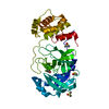
| Structure viewer | Molecule: MolmilJmol/JSmol |
|---|
- Downloads & links
-Download
| PDBx/mmCIF format | 4r62.cif.gz | 57.5 KB | Display | PDBx/mmCIF format |
|---|---|---|---|---|
| PDB format | pdb4r62.ent.gz | 40.3 KB | Display | PDB format |
| PDBx/mmJSON format | 4r62.json.gz | Tree view | PDBx/mmJSON format | |
| Others |  Other downloads Other downloads |
-Validation report
| Arichive directory | https://data.pdbj.org/pub/pdb/validation_reports/r6/4r62ftp://data.pdbj.org/pub/pdb/validation_reports/r6/4r62 | HTTPS FTP |
|---|
-Related structure data
| Related structure data | 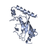 1ayzS  1ubqS  4qwh S: Starting model for refinement |
|---|---|
| Similar structure data |


-Links
 PDBj
PDBj


- Assembly
| Deposited unit | 
| ||||||||
|---|---|---|---|---|---|---|---|---|---|
| 1 |
| ||||||||
| Unit cell |
|
-Components
| #1: Protein | Mass: 19751.699 Da / Num. of mol.: 1 / Fragment: Rad6 / Mutation: C88K Source method: isolated from a genetically manipulated source Source: (gene. exp.) Strain: ATCC 204508 / S288c / Gene: RAD6, UBC2, YGL058W / Plasmid: pMALc2 / Production host:  |
|---|---|
| #2: Protein | Mass: 8818.117 Da / Num. of mol.: 1 / Fragment: Ubiquitin Source method: isolated from a genetically manipulated source Source: (gene. exp.) Homo sapiens (human) / Strain: human / Gene: RPS27A, UBA80, UBCEP1, Ubiquitin / Plasmid: pET3a / Production host: |
| #3: Chemical | ChemComp-ACT /   Mass: 59.044 Da / Num. of mol.: 1 / Source method: obtained synthetically / Formula: C2H3O2 Mass: 59.044 Da / Num. of mol.: 1 / Source method: obtained synthetically / Formula: C2H3O2 |
| #4: Water | ChemComp-HOH /  Mass: 18.015 Da / Num. of mol.: 12 / Source method: isolated from a natural source / Formula: H2O Mass: 18.015 Da / Num. of mol.: 12 / Source method: isolated from a natural source / Formula: H2O |
-Experimental details
-Experiment
| Experiment | Method: X-RAY DIFFRACTION / Number of used crystals: 1 |
|---|
- Sample preparation
| Crystal | Density Matthews: 1.99 Å3/Da / Density % sol: 38.23 % |
|---|---|
| Crystal grow | Temperature: 293 K / Method: vapor diffusion, hanging drop / pH: 5.4 Details: 24% PEG 300, 50 mM sodium acetate, pH 5.4, VAPOR DIFFUSION, HANGING DROP, temperature 293K |
-Data collection
| Diffraction | Mean temperature: 77 K |
|---|---|
| Diffraction source | Source: SYNCHROTRON / Site: APS  / Beamline: 23-ID-D / Wavelength: 1.03 Å / Beamline: 23-ID-D / Wavelength: 1.03 Å |
| Detector | Type: PILATUS3 6M / Detector: PIXEL / Date: Aug 10, 2014 / Details: Silicon sensor |
| Radiation | Monochromator: Silicon sensor / Protocol: SINGLE WAVELENGTH / Monochromatic (M) / Laue (L): M / Scattering type: x-ray |
| Radiation wavelength | Wavelength: 1.03 Å / Relative weight: 1 |
| Reflection | Resolution: 2.2→50 Å / Num. all: 20019 / Num. obs: 11656 / % possible obs: 96.5 % / Redundancy: 2.9 % / Biso Wilson estimate: 59.83 Å2 / Rmerge(I) obs: 0.106 / Rsym value: 0.104 / Net I/σ(I): 2.15 |
- Processing
| Software |
| |||||||||||||||||||||||||||||||||||
|---|---|---|---|---|---|---|---|---|---|---|---|---|---|---|---|---|---|---|---|---|---|---|---|---|---|---|---|---|---|---|---|---|---|---|---|---|
| Refinement | Method to determine structure: MOLECULAR REPLACEMENT Starting model: 1AYZ and 1UBQ Resolution: 2.28→41.107 Å / SU ML: 0.35 / σ(F): 1.34 / Phase error: 34.77 / Stereochemistry target values: ML
| |||||||||||||||||||||||||||||||||||
| Solvent computation | Shrinkage radii: 0.9 Å / VDW probe radii: 1.11 Å / Solvent model: FLAT BULK SOLVENT MODEL | |||||||||||||||||||||||||||||||||||
| Refinement step | Cycle: LAST / Resolution: 2.28→41.107 Å
| |||||||||||||||||||||||||||||||||||
| Refine LS restraints |
| |||||||||||||||||||||||||||||||||||
| LS refinement shell |
|
