[English] 日本語
Yorodumi
- PDB-4r4p: Crystal Structure of the VS ribozyme-A756G mutant -

+
Open data


ID or keywords:

Loading...

-
Basic information

Entry
Database: PDB / ID: 4r4p
TitleCrystal Structure of the VS ribozyme-A756G mutant
ComponentsVS ribozyme RNA
KeywordsRNA / NA / Dimer
Function / homologyRNA / RNA (> 10) / RNA (> 100)
Function and homology information
Biological speciesNEUROSPORA (fungus)
MethodX-RAY DIFFRACTION / SYNCHROTRON / MOLECULAR REPLACEMENT / Resolution: 3.07 Å
AuthorsPiccirilli, J.A. / Suslov, N.B. / Dasgupta, S. / Huang, H. / Lilley, D.M.J. / Rice, P.A.
Citation
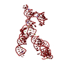
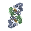
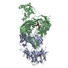
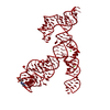
Journal: Nat Chem Biol / Year: 2015
Title: Crystal structure of the Varkud satellite ribozyme.
Authors: Nikolai B Suslov / Saurja DasGupta / Hao Huang / James R Fuller / David M J Lilley / Phoebe A Rice / Joseph A Piccirilli /
Abstract: The Varkud satellite (VS) ribozyme mediates rolling-circle replication of a plasmid found in the Neurospora mitochondrion. We report crystal structures of this ribozyme from Neurospora intermedia at ...The Varkud satellite (VS) ribozyme mediates rolling-circle replication of a plasmid found in the Neurospora mitochondrion. We report crystal structures of this ribozyme from Neurospora intermedia at 3.1 Å resolution, which revealed an intertwined dimer formed by an exchange of substrate helices. In each protomer, an arrangement of three-way helical junctions organizes seven helices into a global fold that creates a docking site for the substrate helix of the other protomer, resulting in the formation of two active sites in trans. This mode of RNA-RNA association resembles the process of domain swapping in proteins and has implications for RNA regulation and evolution. Within each active site, adenine and guanine nucleobases abut the scissile phosphate, poised to serve direct roles in catalysis. Similarities to the active sites of the hairpin and hammerhead ribozymes highlight the functional importance of active-site features, underscore the ability of RNA to access functional architectures from distant regions of sequence space, and suggest convergent evolution.
History
DepositionAug 19, 2014Deposition site: RCSB / Processing site: RCSB
Revision 1.0Sep 30, 2015Provider: repository / Type: Initial release
Revision 1.1Nov 4, 2015Group: Database references
Revision 1.2Sep 20, 2023Group: Data collection / Database references ...Data collection / Database references / Derived calculations / Refinement description
Category: chem_comp_atom / chem_comp_bond ...chem_comp_atom / chem_comp_bond / database_2 / pdbx_initial_refinement_model / pdbx_struct_conn_angle / struct_conn / struct_site
Item: _database_2.pdbx_DOI / _database_2.pdbx_database_accession ..._database_2.pdbx_DOI / _database_2.pdbx_database_accession / _pdbx_struct_conn_angle.ptnr1_auth_seq_id / _pdbx_struct_conn_angle.ptnr3_auth_seq_id / _pdbx_struct_conn_angle.value / _struct_conn.pdbx_dist_value / _struct_conn.pdbx_leaving_atom_flag / _struct_conn.ptnr1_auth_seq_id / _struct_conn.ptnr1_label_asym_id / _struct_conn.ptnr2_auth_seq_id / _struct_site.pdbx_auth_asym_id / _struct_site.pdbx_auth_comp_id / _struct_site.pdbx_auth_seq_id

-
Structure visualization

Structure viewerMolecule:
MolmilJmol/JSmol

Downloads & links

-
Assembly

Deposited unit
A: VS ribozyme RNA
hetero molecules


Theoretical massNumber of molelcules
Total (without water)60,2473
Polymers60,1991
Non-polymers492
Water23413
1
A: VS ribozyme RNA
hetero molecules

A: VS ribozyme RNA
hetero molecules


Theoretical massNumber of molelcules
Total (without water)120,4956
Polymers120,3972
Non-polymers974
Water362
TypeNameSymmetry operationNumber
identity operation1_555x,y,z1
crystal symmetry operation8_555-y,-x,-z+1/21
Buried area6060 Å2
ΔGint-32 kcal/mol
Surface area59980 Å2
MethodPISA
Unit cell
Length a, b, c (Å)102.680, 102.680, 211.021
Angle α, β, γ (deg.)90.00, 90.00, 90.00
Int Tables number96
Space group name H-MP43212

-
Components

#1: RNA chain VS ribozyme RNA


Mass: 60198.699 Da / Num. of mol.: 1 / Mutation: A756G / Source method: obtained synthetically / Details: generated by in-vitro transcription / Source: (synth.) NEUROSPORA (fungus)
#2: Chemical ChemComp-MG / MAGNESIUM ION


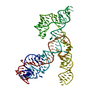
Mass: 24.305 Da / Num. of mol.: 2 / Source method: obtained synthetically / Formula: Mg
#3: Water ChemComp-HOH / water


Mass: 18.015 Da / Num. of mol.: 13 / Source method: isolated from a natural source / Formula: H2O

-
Experimental details

-
Experiment

ExperimentMethod: X-RAY DIFFRACTION / Number of used crystals: 1

-
Sample preparation

CrystalDensity Matthews: 4.62 Å3/Da / Density % sol: 73.38 %
Crystal growTemperature: 293 K / Method: vapor diffusion, hanging drop / pH: 5.8
Details: 0.1 M Sodium citrate.2H20, 2M Ammonium sulfate, 10% 1,3 butanediol, pH 5.8, VAPOR DIFFUSION, HANGING DROP, temperature 293K

-
Data collection

DiffractionMean temperature: 77 K
Diffraction sourceSource: SYNCHROTRON / Site: APS / Beamline: 24-ID-E / Wavelength: 0.97918 Å
DetectorType: ADSC QUANTUM 315 / Detector: CCD / Date: Nov 25, 2013
RadiationMonochromator: Single-crystal side-bounce / Protocol: SINGLE WAVELENGTH / Monochromatic (M) / Laue (L): M / Scattering type: x-ray
Radiation wavelengthWavelength: 0.97918 Å / Relative weight: 1
ReflectionResolution: 3.07→41.469 Å / Num. all: 21134 / Num. obs: 21134 / % possible obs: 96.69 % / Observed criterion σ(F): 0 / Observed criterion σ(I): 2
Reflection shellResolution: 3.07→3.18 Å / % possible all: 96.69

-
Processing

Software
NameVersionClassification
HKL-2000data collection
HKL-2000data reduction
HKL-2000data scaling
PHENIX(phenix.refine: 1.8.2_1309)refinement
SHELXSphasing
RefinementMethod to determine structure: MOLECULAR REPLACEMENT
Starting model: 4R4V
Resolution: 3.07→41.469 Å / SU ML: 0.55 / σ(F): 1.34 / Phase error: 40.32 / Stereochemistry target values: ML
RfactorNum. reflection% reflectionSelection details
Rfree0.269 1872 8.86 %Phenix Protocol
Rwork0.235 ---
all0.2381 21134 --
obs0.235 21134 96.69 %-
Solvent computationShrinkage radii: 0.9 Å / VDW probe radii: 1.11 Å / Solvent model: FLAT BULK SOLVENT MODEL
Refinement stepCycle: LAST / Resolution: 3.07→41.469 Å
ProteinNucleic acidLigandSolventTotal
Num. atoms0 3986 2 13 4001
Refine LS restraints
Refine-IDTypeDev idealNumber
X-RAY DIFFRACTIONf_bond_d0.0094471
X-RAY DIFFRACTIONf_angle_d1.8966984
X-RAY DIFFRACTIONf_dihedral_angle_d16.7942222
X-RAY DIFFRACTIONf_chiral_restr0.076929
X-RAY DIFFRACTIONf_plane_restr0.011186
LS refinement shell
Resolution (Å)Rfactor RfreeNum. reflection RfreeRfactor RworkNum. reflection RworkRefine-ID% reflection obs (%)
3.07-3.1530.43561400.44821451X-RAY DIFFRACTION96
3.153-3.24570.46161410.4221417X-RAY DIFFRACTION95
3.2457-3.35040.43151390.41061417X-RAY DIFFRACTION95
3.3504-3.47010.44221360.38421421X-RAY DIFFRACTION95
3.4701-3.6090.39591340.38921414X-RAY DIFFRACTION94
3.609-3.77310.40291390.35621440X-RAY DIFFRACTION95
3.7731-3.97190.40731410.31461440X-RAY DIFFRACTION95
3.9719-4.22060.31961420.2971460X-RAY DIFFRACTION96
4.2206-4.54610.34231470.2791519X-RAY DIFFRACTION99
4.5461-5.00280.29331490.24371532X-RAY DIFFRACTION100
5.0028-5.72520.24321520.22441551X-RAY DIFFRACTION100
5.7252-7.20690.2311530.19791575X-RAY DIFFRACTION100
7.2069-41.47280.14971590.12141625X-RAY DIFFRACTION97

+
About Yorodumi

-
News

-
Feb 9, 2022. New format data for meta-information of EMDB entries

New format data for meta-information of EMDB entries

  • Version 3 of the EMDB header file is now the official format.
  • The previous official version 1.9 will be removed from the archive.

Related info.:EMDB header

External links:wwPDB to switch to version 3 of the EMDB data model

-
Aug 12, 2020. Covid-19 info

Covid-19 info

URL: https://pdbjlvh1.pdbj.org/emnavi/covid19.php

New page: Covid-19 featured information page in EM Navigator.

Related info.:Covid-19 info / Mar 5, 2020. Novel coronavirus structure data

+
Mar 5, 2020. Novel coronavirus structure data

Novel coronavirus structure data

Related info.:Yorodumi Speices / Aug 12, 2020. Covid-19 info

External links:COVID-19 featured content - PDBj / Molecule of the Month (242):Coronavirus Proteases

+
Jan 31, 2019. EMDB accession codes are about to change! (news from PDBe EMDB page)

EMDB accession codes are about to change! (news from PDBe EMDB page)

  • The allocation of 4 digits for EMDB accession codes will soon come to an end. Whilst these codes will remain in use, new EMDB accession codes will include an additional digit and will expand incrementally as the available range of codes is exhausted. The current 4-digit format prefixed with “EMD-” (i.e. EMD-XXXX) will advance to a 5-digit format (i.e. EMD-XXXXX), and so on. It is currently estimated that the 4-digit codes will be depleted around Spring 2019, at which point the 5-digit format will come into force.
  • The EM Navigator/Yorodumi systems omit the EMD- prefix.

Related info.:Q: What is EMD? / ID/Accession-code notation in Yorodumi/EM Navigator

External links:EMDB Accession Codes are Changing Soon! / Contact to PDBj

+
Jul 12, 2017. Major update of PDB

Major update of PDB

  • wwPDB released updated PDB data conforming to the new PDBx/mmCIF dictionary.
  • This is a major update changing the version number from 4 to 5, and with Remediation, in which all the entries are updated.
  • In this update, many items about electron microscopy experimental information are reorganized (e.g. em_software).
  • Now, EM Navigator and Yorodumi are based on the updated data.

External links:wwPDB Remediation / Enriched Model Files Conforming to OneDep Data Standards Now Available in the PDB FTP Archive

-
Yorodumi

Thousand views of thousand structures

  • Yorodumi is a browser for structure data from EMDB, PDB, SASBDB, etc.
  • This page is also the successor to EM Navigator detail page, and also detail information page/front-end page for Omokage search.
  • The word "yorodu" (or yorozu) is an old Japanese word meaning "ten thousand". "mi" (miru) is to see.

Related info.:EMDB / PDB / SASBDB / Comparison of 3 databanks / Yorodumi Search / Aug 31, 2016. New EM Navigator & Yorodumi / Yorodumi Papers / Jmol/JSmol / Function and homology information / Changes in new EM Navigator and Yorodumi

Read more