 Movie
Movie Controller

[English] 日本語
 Yorodumi
- PDB-4r0p: Ifqins, an amyloid forming segment from human lysozyme spanning r... -
+ Open data

- Basic information
| Entry | Database: PDB / ID: 4r0p | ||||||
|---|---|---|---|---|---|---|---|
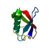
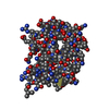
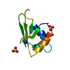
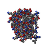
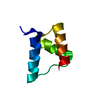
| Title | Ifqins, an amyloid forming segment from human lysozyme spanning residues 56-61 | ||||||
 Components Components | Lysozyme C | ||||||
 Keywords Keywords | PROTEIN FIBRIL / amyloid-like protofibril | ||||||
| Function / homology |  Function and homology information Function and homology informationantimicrobial humoral response / Antimicrobial peptides / specific granule lumen / lysozyme / lysozyme activity / azurophil granule lumen / tertiary granule lumen / killing of cells of another organism / defense response to Gram-negative bacterium / defense response to bacterium ...antimicrobial humoral response / Antimicrobial peptides / specific granule lumen / lysozyme / lysozyme activity / azurophil granule lumen / tertiary granule lumen / killing of cells of another organism / defense response to Gram-negative bacterium / defense response to bacterium / defense response to Gram-positive bacterium / Amyloid fiber formation / inflammatory response / Neutrophil degranulation / : / extracellular exosome / extracellular region / identical protein binding Similarity search - Function | ||||||
| Biological species |  Homo sapiens (human) Homo sapiens (human) | ||||||
| Method |  X-RAY DIFFRACTION / SYNCHROTRON / MOLECULAR REPLACEMENT / molecular replacement / Resolution: 1.52 Å X-RAY DIFFRACTION / SYNCHROTRON / MOLECULAR REPLACEMENT / molecular replacement / Resolution: 1.52 Å | ||||||
 Authors Authors | Sievers, S. / Eisenberg, D.S. / Sawaya, M.R. | ||||||
 Citation Citation | Journal: J.Am.Chem.Soc. / Year: 2014 Title: Structure-based design of functional amyloid materials. Authors: Li, D. / Jones, E.M. / Sawaya, M.R. / Furukawa, H. / Luo, F. / Ivanova, M. / Sievers, S.A. / Wang, W. / Yaghi, O.M. / Liu, C. / Eisenberg, D.S. | ||||||
| History |
|
- Structure visualization
| Structure viewer | Molecule: MolmilJmol/JSmol |
|---|
- Downloads & links
-Download
| PDBx/mmCIF format | 4r0p.cif.gz | 9.2 KB | Display | PDBx/mmCIF format |
|---|---|---|---|---|
| PDB format | pdb4r0p.ent.gz | 5.5 KB | Display | PDB format |
| PDBx/mmJSON format | 4r0p.json.gz | Tree view | PDBx/mmJSON format | |
| Others |  Other downloads Other downloads |
-Validation report
| Arichive directory | https://data.pdbj.org/pub/pdb/validation_reports/r0/4r0pftp://data.pdbj.org/pub/pdb/validation_reports/r0/4r0p | HTTPS FTP |
|---|
-Related structure data


-Links
 PDBj
PDBj

- Assembly
| Deposited unit | 
| ||||||||
|---|---|---|---|---|---|---|---|---|---|
| 1 | x 10
| ||||||||
| Unit cell |
| ||||||||
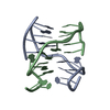
| Details | There are two choices of the biological unit (fibrils). The first choice is a pair of indefinitely long beta sheets constructed from chain A and unit cell translations along the b direction (the b direction corresponds to the fiber axis) (i.e. X,Y,Z; X,Y+1,Z; X,Y+2,Z; etc.) together with a symmetry related sheet formed from -X,1/2+Y,-1/2-Z and its unit cell translations along the b direction (i.e. -X,3/2+Y,-1/2-Z; -X,5/2+Y,-1/2-Z, etc.). The second choice of biological unit is a pair of beta sheets constructed from chain A and unit cell translations along the b direction (i.e. X,Y,Z; X,Y+1,Z; X,Y+2,Z; etc.) together with a symmetry related sheet formed from -1-X,1/2+Y,-1/2-Z and its unit cell translations along the b direction (i.e. -1-X,3/2+Y,-1/2-Z; -1-X,5/2+Y,-1/2-Z, etc.). REMARK 350 displays 5 strands from both sheets for the second choice. |
-Components
| #1: Protein/peptide | Mass: 720.814 Da / Num. of mol.: 1 / Fragment: UNP RESIDUES 74-79 / Source method: obtained synthetically Details: IFQINS hexapeptide (residues 56-61) from human lysozyme, synthesized Source: (synth.) Homo sapiens (human) / References: UniProt: P61626, lysozyme |
|---|---|
| #2: Water | ChemComp-HOH /  Mass: 18.015 Da / Num. of mol.: 2 / Source method: isolated from a natural source / Formula: H2O Mass: 18.015 Da / Num. of mol.: 2 / Source method: isolated from a natural source / Formula: H2O |
-Experimental details
-Experiment
| Experiment | Method: X-RAY DIFFRACTION / Number of used crystals: 1 |
|---|
- Sample preparation
| Crystal | Density Matthews: 1.42 Å3/Da / Density % sol: 13.51 % / Mosaicity: 0.3 ° |
|---|---|
| Crystal grow | Temperature: 298 K / Method: vapor diffusion, hanging drop / pH: 6.5 Details: reservoir contained 0.1M Bis-Tris pH 6.5, 3.0M Sodium Chloride, vapor diffusion, hanging drop, temperature 298K |
-Data collection
| Diffraction | Mean temperature: 100 K | ||||||||||||||||||||||||||||||||||||
|---|---|---|---|---|---|---|---|---|---|---|---|---|---|---|---|---|---|---|---|---|---|---|---|---|---|---|---|---|---|---|---|---|---|---|---|---|---|
| Diffraction source | Source: SYNCHROTRON / Site: ESRF  / Beamline: ID13 / Wavelength: 0.9466 Å / Beamline: ID13 / Wavelength: 0.9466 Å | ||||||||||||||||||||||||||||||||||||
| Detector | Type: MAR CCD 165 mm / Detector: CCD / Date: May 16, 2006 | ||||||||||||||||||||||||||||||||||||
| Radiation | Protocol: SINGLE WAVELENGTH / Monochromatic (M) / Laue (L): M / Scattering type: x-ray | ||||||||||||||||||||||||||||||||||||
| Radiation wavelength | Wavelength: 0.9466 Å / Relative weight: 1 | ||||||||||||||||||||||||||||||||||||
| Reflection | Resolution: 1.5→90 Å / Num. all: 753 / Num. obs: 753 / % possible obs: 93.8 % / Observed criterion σ(I): -3 / Redundancy: 13.1 % / Biso Wilson estimate: 14.5 Å2 / Rmerge(I) obs: 0.181 / Χ2: 1.055 / Net I/σ(I): 5.8 | ||||||||||||||||||||||||||||||||||||
| Reflection shell | Diffraction-ID: 1 / Rejects: _
|
-Phasing
| Phasing | Method: molecular replacement | |||||||||
|---|---|---|---|---|---|---|---|---|---|---|
| Phasing MR | Model details: Phaser MODE: MR_AUTO
|
- Processing
| Software |
| |||||||||||||||||||||||||||||||||||||||||||||||||||||||||||||||||||||||||||||||||||||
|---|---|---|---|---|---|---|---|---|---|---|---|---|---|---|---|---|---|---|---|---|---|---|---|---|---|---|---|---|---|---|---|---|---|---|---|---|---|---|---|---|---|---|---|---|---|---|---|---|---|---|---|---|---|---|---|---|---|---|---|---|---|---|---|---|---|---|---|---|---|---|---|---|---|---|---|---|---|---|---|---|---|---|---|---|---|---|
| Refinement | Method to determine structure: MOLECULAR REPLACEMENT / Resolution: 1.52→21.61 Å / Cor.coef. Fo:Fc: 0.969 / Cor.coef. Fo:Fc free: 0.966 / WRfactor Rfree: 0.1884 / WRfactor Rwork: 0.1458 / FOM work R set: 0.8701 / SU B: 1.566 / SU ML: 0.054 / SU R Cruickshank DPI: 0.0948 / SU Rfree: 0.0969 / Cross valid method: THROUGHOUT / σ(F): 0 / ESU R: 0.095 / ESU R Free: 0.097 / Stereochemistry target values: MAXIMUM LIKELIHOOD / Details: HYDROGENS HAVE BEEN ADDED IN THE RIDING POSITIONS
| |||||||||||||||||||||||||||||||||||||||||||||||||||||||||||||||||||||||||||||||||||||
| Solvent computation | Ion probe radii: 0.8 Å / Shrinkage radii: 0.8 Å / VDW probe radii: 1.4 Å / Solvent model: MASK | |||||||||||||||||||||||||||||||||||||||||||||||||||||||||||||||||||||||||||||||||||||
| Displacement parameters | Biso max: 20.85 Å2 / Biso mean: 4.924 Å2 / Biso min: 2 Å2
| |||||||||||||||||||||||||||||||||||||||||||||||||||||||||||||||||||||||||||||||||||||
| Refinement step | Cycle: LAST / Resolution: 1.52→21.61 Å /
| |||||||||||||||||||||||||||||||||||||||||||||||||||||||||||||||||||||||||||||||||||||
| Refine LS restraints |
| |||||||||||||||||||||||||||||||||||||||||||||||||||||||||||||||||||||||||||||||||||||
| LS refinement shell | Resolution: 1.524→1.564 Å / Total num. of bins used: 20
|
