Mass: 18.015 Da / Num. of mol.: 4 / Source method: isolated from a natural source / Formula: H2O
Has protein modification
Y
Sequence details
SEQUENCE THE CONSTRUCT (RESIDUES 1-264) WAS EXPRESSED WITH A PURIFICATION TAG MGSDKIHHHHHHENLYFQG. ...SEQUENCE THE CONSTRUCT (RESIDUES 1-264) WAS EXPRESSED WITH A PURIFICATION TAG MGSDKIHHHHHHENLYFQG. THE TAG WAS REMOVED WITH TEV PROTEASE LEAVING ONLY A GLYCINE (0) FOLLOWED BY THE TARGET SEQUENCE.
-
Experimental details
-
Experiment
Experiment
Method: X-RAY DIFFRACTION / Number of used crystals: 1
-
Sample preparation
Crystal
Density Matthews: 2.31 Å3/Da / Density % sol: 46.75 %
Crystal grow
Temperature: 293 K / Method: vapor diffusion, sitting drop / pH: 7.47 Details: 53.00% polyethylene glycol 200, 0.1M HEPES pH 7.47, NANODROP, VAPOR DIFFUSION, SITTING DROP, temperature 293K
Resolution: 2.65→29.944 Å / Num. all: 8992 / Num. obs: 8992 / % possible obs: 99.9 % / Redundancy: 11.6 % / Biso Wilson estimate: 86.177 Å2 / Rsym value: 0.086 / Net I/σ(I): 16.2
Reflection shell
Diffraction-ID: 1
Resolution (Å)
Redundancy (%)
Mean I/σ(I) obs
Num. measured all
Num. unique all
Rsym value
% possible all
Rmerge(I) obs
2.65-2.72
12.4
2.3
8051
648
1.267
100
2.72-2.79
12.4
0.9
7711
621
0.886
100
0.013
2.79-2.87
12.4
1.1
7503
607
0.679
100
0.013
2.87-2.96
12
1.5
7295
606
0.5
100
0.013
2.96-3.06
11.6
1.9
6717
577
0.399
100
0.013
3.06-3.17
11.1
2.5
6288
564
0.307
99.9
0.013
3.17-3.29
10.5
3.7
5665
542
0.2
100
0.013
3.29-3.42
12.3
4.6
6511
531
0.163
100
0.013
3.42-3.57
12.2
5.3
6051
494
0.131
100
0.013
3.57-3.75
11.8
6.6
5808
491
0.101
100
0.013
3.75-3.95
11.7
7.6
5422
465
0.089
100
0.013
3.95-4.19
10.7
9
4695
440
0.07
100
0.013
4.19-4.48
11.3
10.1
4679
414
0.062
100
0.013
4.48-4.84
12.2
10
4826
395
0.06
100
0.013
4.84-5.3
11.8
9.9
4270
361
0.063
100
0.013
5.3-5.93
11
9.8
3730
339
0.064
100
0.013
5.93-6.84
9.9
9.8
2949
298
0.062
100
0.013
6.84-8.38
11.6
9.6
3004
260
0.063
99.9
0.013
8.38-11.85
9.9
11.2
2100
213
0.051
99.9
0.013
11.85-29.944
9.2
9.1
1161
126
0.054
93.8
0.013
-
Phasing
Phasing
Method: MAD
-
Processing
Software
Name
Version
Classification
NB
MolProbity
3beta29
modelbuilding
PDB_EXTRACT
3.1
dataextraction
SHELX
phasing
SHARP
phasing
SCALA
3.3.20
datascaling
BUSTER-TNT
2.10.0
refinement
MOSFLM
datareduction
SHELXD
phasing
BUSTER
2.10.0
refinement
Refinement
Method to determine structure: MAD / Resolution: 2.65→29.944 Å / Cor.coef. Fo:Fc: 0.9372 / Cor.coef. Fo:Fc free: 0.9132 / Occupancy max: 1 / Occupancy min: 0.75 / Cross valid method: THROUGHOUT / σ(F): 0 Details: 1. A MET-INHIBITION PROTOCOL WAS USED FOR SELENOMETHIONINE INCORPORATION DURING PROTEIN EXPRESSION. THE OCCUPANCY OF THE SE ATOMS IN THE MSE RESIDUES WAS REDUCED TO 0.75 FOR THE REDUCED ...Details: 1. A MET-INHIBITION PROTOCOL WAS USED FOR SELENOMETHIONINE INCORPORATION DURING PROTEIN EXPRESSION. THE OCCUPANCY OF THE SE ATOMS IN THE MSE RESIDUES WAS REDUCED TO 0.75 FOR THE REDUCED SCATTERING POWER DUE TO PARTIAL S-MET INCORPORATION. 2. ATOM RECORD CONTAINS SUM OF TLS AND RESIDUAL B FACTORS. ANISOU RECORD CONTAINS SUM OF TLS AND RESIDUAL U FACTORS. 3. THE MAD PHASES WERE USED AS RESTRAINTS DURING REFINEMENT. 4. POLYETHYLENE GLYCOL 200 FRAGMENT (PG4) FROM THE CRYSTALLIZATION WERE MODELED INTO THE STRUCTURE. 5. THE MODELING OF ZINC (ZN) INTO THE STRUCTURE IS SUPPORTED BY ANOMALOUS DIFFERENCE DENSITY. 6. SODIUM (NA) FROM PURIFICATION BUFFER WAS MODELED INTO THE STRUCTURE.
In the structure databanks used in Yorodumi, some data are registered as the other names, "COVID-19 virus" and "2019-nCoV". Here are the details of the virus and the list of structure data.
Jan 31, 2019. EMDB accession codes are about to change! (news from PDBe EMDB page)
EMDB accession codes are about to change! (news from PDBe EMDB page)
The allocation of 4 digits for EMDB accession codes will soon come to an end. Whilst these codes will remain in use, new EMDB accession codes will include an additional digit and will expand incrementally as the available range of codes is exhausted. The current 4-digit format prefixed with “EMD-” (i.e. EMD-XXXX) will advance to a 5-digit format (i.e. EMD-XXXXX), and so on. It is currently estimated that the 4-digit codes will be depleted around Spring 2019, at which point the 5-digit format will come into force.
The EM Navigator/Yorodumi systems omit the EMD- prefix.
Related info.:Q: What is EMD? / ID/Accession-code notation in Yorodumi/EM Navigator
Yorodumi is a browser for structure data from EMDB, PDB, SASBDB, etc.
This page is also the successor to EM Navigator detail page, and also detail information page/front-end page for Omokage search.
The word "yorodu" (or yorozu) is an old Japanese word meaning "ten thousand". "mi" (miru) is to see.
Related info.:EMDB / PDB / SASBDB / Comparison of 3 databanks / Yorodumi Search / Aug 31, 2016. New EM Navigator & Yorodumi / Yorodumi Papers / Jmol/JSmol / Function and homology information / Changes in new EM Navigator and Yorodumi
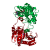
 Movie
Movie Controller

 Yorodumi
 Open data
 Basic information
 Components
 Keywords
 Function and homology information
 X-RAY DIFFRACTION /
  Authors
 Citation
 Structure visualization
 Downloads & links
 Other downloads

 PDBj
PDBj


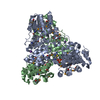
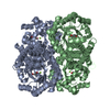
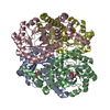
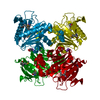
 Assembly

 Mass: 65.409 Da / Num. of mol.: 1 / Source method: obtained synthetically / Formula: Zn
 Mass: 194.226 Da / Num. of mol.: 1 / Source method: obtained synthetically / Formula: C8H18O5 / Comment: precipitant*YM
 Mass: 22.990 Da / Num. of mol.: 1 / Source method: obtained synthetically / Formula: Na
 Mass: 18.015 Da / Num. of mol.: 4 / Source method: isolated from a natural source / Formula: H2O
 Sample preparation
 / Beamline: BL12-2 / Wavelength: 0.9116,0.9794
 Processing