[English] 日本語
Yorodumi
- PDB-4qsm: Crystal structure of human muscle L-lactate dehydrogenase in comp... -

+
Open data


ID or keywords:

Loading...

-
Basic information

Entry
Database: PDB / ID: 4qsm
Title
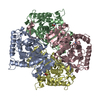
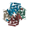
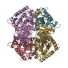
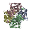
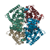
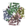
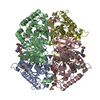
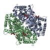
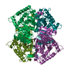
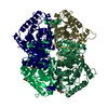
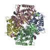
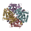
Crystal structure of human muscle L-lactate dehydrogenase in complex with inhibitor 2, 3-{[7-(2,4-dimethoxypyrimidin-5-yl)-3-sulfamoylquinolin-4-yl]amino}benzoic acid
ComponentsL-lactate dehydrogenase A chain
Keywordsoxidoreductase/oxidoreductase Inhibitor / Rossmann Fold / oxidoreductase-oxidoreductase Inhibitor complex
Function / homology
Function and homology information


oxidoreductase complex / Pyruvate metabolism / L-lactate dehydrogenase / lactate metabolic process / L-lactate dehydrogenase activity / pyruvate metabolic process / substantia nigra development / glycolytic process / cadherin binding / mitochondrion ...oxidoreductase complex / Pyruvate metabolism / L-lactate dehydrogenase / lactate metabolic process / L-lactate dehydrogenase activity / pyruvate metabolic process / substantia nigra development / glycolytic process / cadherin binding / mitochondrion / extracellular exosome / identical protein binding / membrane / nucleus / cytosol
Similarity search - Function
L-lactate dehydrogenase / L-lactate dehydrogenase active site. / L-lactate dehydrogenase, active site / L-2-Hydroxyisocaproate Dehydrogenase; Chain A, domain 2 / Lactate dehydrogenase/glycoside hydrolase, family 4, C-terminal / L-lactate/malate dehydrogenase / Lactate/malate dehydrogenase, N-terminal / Lactate/malate dehydrogenase, C-terminal / lactate/malate dehydrogenase, NAD binding domain / lactate/malate dehydrogenase, alpha/beta C-terminal domain ...L-lactate dehydrogenase / L-lactate dehydrogenase active site. / L-lactate dehydrogenase, active site / L-2-Hydroxyisocaproate Dehydrogenase; Chain A, domain 2 / Lactate dehydrogenase/glycoside hydrolase, family 4, C-terminal / L-lactate/malate dehydrogenase / Lactate/malate dehydrogenase, N-terminal / Lactate/malate dehydrogenase, C-terminal / lactate/malate dehydrogenase, NAD binding domain / lactate/malate dehydrogenase, alpha/beta C-terminal domain / Lactate dehydrogenase/glycoside hydrolase, family 4, C-terminal / NAD(P)-binding Rossmann-like Domain / NAD(P)-binding domain superfamily / Alpha-Beta Complex / Rossmann fold / 3-Layer(aba) Sandwich / Alpha Beta
Similarity search - Domain/homology
Chem-38K / L-lactate dehydrogenase A chain
Similarity search - Component
Biological speciesHomo sapiens (human)
MethodX-RAY DIFFRACTION / SYNCHROTRON / MOLECULAR REPLACEMENT / Resolution: 3 Å
AuthorsKolappan, S. / Craig, L.
CitationJournal: Acta Crystallogr.,Sect.D / Year: 2015
Title: Structures of lactate dehydrogenase A (LDHA) in apo, ternary and inhibitor-bound forms.
Authors: Kolappan, S. / Shen, D.L. / Mosi, R. / Sun, J. / McEachern, E.J. / Vocadlo, D.J. / Craig, L.
History
DepositionJul 6, 2014Deposition site: RCSB / Processing site: RCSB
Revision 1.0Dec 3, 2014Provider: repository / Type: Initial release
Revision 1.1Feb 25, 2015Group: Database references
Revision 1.2Nov 22, 2017Group: Refinement description / Category: software
Revision 1.3Feb 28, 2024Group: Data collection / Database references / Derived calculations
Category: chem_comp_atom / chem_comp_bond ...chem_comp_atom / chem_comp_bond / database_2 / struct_ref_seq_dif / struct_site
Item: _database_2.pdbx_DOI / _database_2.pdbx_database_accession ..._database_2.pdbx_DOI / _database_2.pdbx_database_accession / _struct_ref_seq_dif.details / _struct_site.pdbx_auth_asym_id / _struct_site.pdbx_auth_comp_id / _struct_site.pdbx_auth_seq_id

-
Structure visualization

Structure viewerMolecule:
MolmilJmol/JSmol

Downloads & links

-
Assembly

Deposited unit
A: L-lactate dehydrogenase A chain
B: L-lactate dehydrogenase A chain
C: L-lactate dehydrogenase A chain
D: L-lactate dehydrogenase A chain
E: L-lactate dehydrogenase A chain
F: L-lactate dehydrogenase A chain
G: L-lactate dehydrogenase A chain
H: L-lactate dehydrogenase A chain
hetero molecules


Theoretical massNumber of molelcules
Total (without water)303,31116
Polymers299,4598
Non-polymers3,8528
Water1,08160
1
A: L-lactate dehydrogenase A chain
B: L-lactate dehydrogenase A chain
C: L-lactate dehydrogenase A chain
D: L-lactate dehydrogenase A chain
hetero molecules


Theoretical massNumber of molelcules
Total (without water)151,6558
Polymers149,7294
Non-polymers1,9264
Water724
TypeNameSymmetry operationNumber
identity operation1_555x,y,z1
Buried area21350 Å2
ΔGint-131 kcal/mol
Surface area47170 Å2
MethodPISA
2
E: L-lactate dehydrogenase A chain
F: L-lactate dehydrogenase A chain
G: L-lactate dehydrogenase A chain
H: L-lactate dehydrogenase A chain
hetero molecules


Theoretical massNumber of molelcules
Total (without water)151,6558
Polymers149,7294
Non-polymers1,9264
Water724
TypeNameSymmetry operationNumber
identity operation1_555x,y,z1
Buried area21130 Å2
ΔGint-138 kcal/mol
Surface area47150 Å2
MethodPISA
Unit cell
Length a, b, c (Å)147.310, 147.310, 334.490
Angle α, β, γ (deg.)90.00, 90.00, 120.00
Int Tables number152
Space group name H-MP3121

-
Components

#1: Protein
L-lactate dehydrogenase A chain / LDH-A / Cell proliferation-inducing gene 19 protein / LDH muscle subunit / LDH-M / Renal carcinoma ...LDH-A / Cell proliferation-inducing gene 19 protein / LDH muscle subunit / LDH-M / Renal carcinoma antigen NY-REN-59


Mass: 37432.344 Da / Num. of mol.: 8
Source method: isolated from a genetically manipulated source
Source: (gene. exp.) Homo sapiens (human) / Gene: LDHA, PIG19 / Production host: Escherichia coli (E. coli) / References: UniProt: P00338, L-lactate dehydrogenase
#2: Chemical
ChemComp-38K / 3-{[7-(2,4-dimethoxypyrimidin-5-yl)-3-sulfamoylquinolin-4-yl]amino}benzoic acid


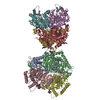
Mass: 481.481 Da / Num. of mol.: 8 / Source method: obtained synthetically / Formula: C22H19N5O6S
#3: Water ChemComp-HOH / water


Mass: 18.015 Da / Num. of mol.: 60 / Source method: isolated from a natural source / Formula: H2O

-
Experimental details

-
Experiment

ExperimentMethod: X-RAY DIFFRACTION / Number of used crystals: 1

-
Sample preparation

CrystalDensity Matthews: 3.5 Å3/Da / Density % sol: 64.84 %
Crystal growTemperature: 293 K / Method: vapor diffusion / pH: 7
Details: 50 mM LiCl, PEG400 41 % w/v, 100 mM Bis-tris propane, pH 7.0, VAPOR DIFFUSION, temperature 293K

-
Data collection

DiffractionMean temperature: 100 K
Diffraction sourceSource: SYNCHROTRON / Site: SSRL / Beamline: BL7-1 / Wavelength: 1 Å
DetectorType: ADSC QUANTUM 315r / Detector: CCD / Date: Jul 2, 2013 / Details: mirrors
RadiationMonochromator: Si(111) / Protocol: SINGLE WAVELENGTH / Monochromatic (M) / Laue (L): M / Scattering type: x-ray
Radiation wavelengthWavelength: 1 Å / Relative weight: 1
ReflectionResolution: 3→35 Å / Num. all: 85010 / Num. obs: 84925 / % possible obs: 99.9 % / Observed criterion σ(F): 2 / Observed criterion σ(I): 2
Reflection shell
Resolution (Å)Redundancy (%)Rmerge(I) obsMean I/σ(I) obsNum. unique allDiffraction-ID% possible all
3.03-3.087.51.022.24419199.9
3.08-3.147.50.8782.54400199.9
3.14-3.217.50.7932.84260199.9
3.21-3.277.50.6723.24204199.9
3.27-3.357.50.534.14125199.9
3.35-3.427.50.4514.84048199.9
3.42-3.517.50.3925.63922199.9
3.51-3.597.50.3346.53887199.9
3.59-3.697.50.2957.33778199.9
3.69-3.797.50.2458.73651199.9
3.79-3.917.50.211103605199.9
3.91-4.037.50.17611.83465199.8
4.03-4.177.40.15213.23376199.8
4.17-4.327.40.1216.33274199.8
4.32-4.497.40.09520.23139199.8
4.49-4.687.40.081233073199.8
4.68-4.97.40.077242875199.7
4.9-5.157.30.07424.22809199.7
5.15-5.447.20.07922.82678199.6
5.44-5.87.20.08820.12535199.6
5.8-6.237.10.07821.52370199.5
6.23-6.777.40.06425.62248199.4
6.77-7.487.40.05926.82072199.3
7.48-8.497.30.04136.71875199
8.49-10.047.20.03148.71678198.6
10.04-12.967.10.02459.41426197.9
12.96-22.456.90.024561135196.2
22.45-3560.02546.8598189.3

-
Processing

Software
NameVersionClassification
Blu-Icedata collection
REFMAC5.7.0029refinement
XDSdata reduction
XSCALEdata scaling
RefinementMethod to determine structure: MOLECULAR REPLACEMENT / Resolution: 3→34.62 Å / Cor.coef. Fo:Fc: 0.955 / Cor.coef. Fo:Fc free: 0.873 / SU B: 30.17 / SU ML: 0.251 / Cross valid method: THROUGHOUT / ESU R Free: 0.387 / Stereochemistry target values: MAXIMUM LIKELIHOOD / Details: HYDROGENS HAVE BEEN ADDED IN THE RIDING POSITIONS
RfactorNum. reflection% reflectionSelection details
Rfree0.247 4518 5.3 %RANDOM
Rwork0.167 ---
obs0.172 80339 99.88 %-
all-84925 --
Solvent computationIon probe radii: 0.8 Å / Shrinkage radii: 0.8 Å / VDW probe radii: 1.2 Å / Solvent model: MASK
Displacement parametersBiso mean: 65.878 Å2
Baniso -1Baniso -2Baniso -3
1--1.38 Å2-1.38 Å20 Å2
2---1.38 Å20 Å2
3---4.48 Å2
Refinement stepCycle: LAST / Resolution: 3→34.62 Å
ProteinNucleic acidLigandSolventTotal
Num. atoms20566 0 272 60 20898
Refine LS restraints
Refine-IDTypeDev idealDev ideal targetNumber
X-RAY DIFFRACTIONr_bond_refined_d0.0110.01921494
X-RAY DIFFRACTIONr_bond_other_d0.0020.0221311
X-RAY DIFFRACTIONr_angle_refined_deg1.5511.98629149
X-RAY DIFFRACTIONr_angle_other_deg0.839349109
X-RAY DIFFRACTIONr_dihedral_angle_1_deg6.43952701
X-RAY DIFFRACTIONr_dihedral_angle_2_deg38.6225.114833
X-RAY DIFFRACTIONr_dihedral_angle_3_deg18.922153979
X-RAY DIFFRACTIONr_dihedral_angle_4_deg20.1421590
X-RAY DIFFRACTIONr_chiral_restr0.0780.23361
X-RAY DIFFRACTIONr_gen_planes_refined0.0070.0223987
X-RAY DIFFRACTIONr_gen_planes_other0.0070.024573
X-RAY DIFFRACTIONr_nbd_refined
X-RAY DIFFRACTIONr_nbd_other
X-RAY DIFFRACTIONr_nbtor_refined
X-RAY DIFFRACTIONr_nbtor_other
X-RAY DIFFRACTIONr_xyhbond_nbd_refined
X-RAY DIFFRACTIONr_xyhbond_nbd_other
X-RAY DIFFRACTIONr_metal_ion_refined
X-RAY DIFFRACTIONr_metal_ion_other
X-RAY DIFFRACTIONr_symmetry_vdw_refined
X-RAY DIFFRACTIONr_symmetry_vdw_other
X-RAY DIFFRACTIONr_symmetry_hbond_refined
X-RAY DIFFRACTIONr_symmetry_hbond_other
X-RAY DIFFRACTIONr_symmetry_metal_ion_refined
X-RAY DIFFRACTIONr_symmetry_metal_ion_other
X-RAY DIFFRACTIONr_mcbond_it
X-RAY DIFFRACTIONr_mcbond_other
X-RAY DIFFRACTIONr_mcangle_it
X-RAY DIFFRACTIONr_mcangle_other
X-RAY DIFFRACTIONr_scbond_it
X-RAY DIFFRACTIONr_scbond_other
X-RAY DIFFRACTIONr_scangle_it
X-RAY DIFFRACTIONr_scangle_other
X-RAY DIFFRACTIONr_long_range_B_refined
X-RAY DIFFRACTIONr_long_range_B_other
X-RAY DIFFRACTIONr_rigid_bond_restr
X-RAY DIFFRACTIONr_sphericity_free
X-RAY DIFFRACTIONr_sphericity_bonded
LS refinement shellResolution: 3→3.077 Å / Total num. of bins used: 20
RfactorNum. reflection% reflection
Rfree0.335 308 -
Rwork0.261 5836 -
obs--99.97 %
Refinement TLS params.

Method: refined / Refine-ID: X-RAY DIFFRACTION

IDL112)L122)L132)L222)L232)L332)S11 (Å °)S12 (Å °)S13 (Å °)S21 (Å °)S22 (Å °)S23 (Å °)S31 (Å °)S32 (Å °)S33 (Å °)T112)T122)T132)T222)T232)T332)Origin x (Å)Origin y (Å)Origin z (Å)
11.5691-0.08770.01611.4098-0.0291.15860.0010.2525-0.2719-0.2235-0.0102-0.12560.1430.10440.00920.2481-0.07010.00590.2092-0.0950.090570.7292-23.3529-29.3494
22.05520.0121-0.12292.2754-0.15880.4561-0.01440.26110.1128-0.3079-0.02650.5213-0.098-0.22380.04090.2559-0.0528-0.0940.2189-0.03750.143145.0715-2.7991-22.985
31.12390.2762-0.43991.79280.16111.99890.0128-0.1518-0.06980.452-0.0392-0.10450.02080.10280.02640.3076-0.1176-0.08490.09920.04310.030169.784-12.55566.6343
42.05160.59220.35922.0974-0.70531.0192-0.0440.0860.23750.0443-0.0409-0.194-0.22040.04210.08480.2649-0.1019-0.03960.1450.02550.08478.19612.467-13.698
52.56080.2539-0.35221.5936-0.12711.4537-0.09370.69530.0176-0.56910.11380.3333-0.001-0.2157-0.02020.4907-0.0951-0.19610.46720.01710.092110.4238-53.0431-80.446
61.8010.5471-0.17482.71730.08651.0665-0.0217-0.1192-0.15920.088-0.03350.24120.0758-0.15220.05510.2531-0.0878-0.05590.2517-0.00580.074110.8947-69.558-51.2789
72.27860.1527-0.06281.33520.05131.02750.01490.16180.2418-0.18920.1027-0.1669-0.03550.151-0.11760.2435-0.04890.00410.1638-0.03760.072641.7088-46.5681-60.9376
81.91180.2635-0.21321.8377-0.02541.05670.0048-0.21910.62560.151-0.00910.2451-0.1855-0.19880.00430.27430.0059-0.0750.2597-0.10350.263514.7886-32.5634-46.4347
Refinement TLS group
IDRefine-IDRefine TLS-IDAuth asym-IDAuth seq-ID
1X-RAY DIFFRACTION1A2 - 333
2X-RAY DIFFRACTION2B2 - 332
3X-RAY DIFFRACTION3C2 - 333
4X-RAY DIFFRACTION4D2 - 333
5X-RAY DIFFRACTION5E2 - 332
6X-RAY DIFFRACTION6F2 - 332
7X-RAY DIFFRACTION7G2 - 332
8X-RAY DIFFRACTION8H2 - 332

+
About Yorodumi

-
News

-
Feb 9, 2022. New format data for meta-information of EMDB entries

New format data for meta-information of EMDB entries

  • Version 3 of the EMDB header file is now the official format.
  • The previous official version 1.9 will be removed from the archive.

Related info.:EMDB header

External links:wwPDB to switch to version 3 of the EMDB data model

-
Aug 12, 2020. Covid-19 info

Covid-19 info

URL: https://pdbjlvh1.pdbj.org/emnavi/covid19.php

New page: Covid-19 featured information page in EM Navigator.

Related info.:Covid-19 info / Mar 5, 2020. Novel coronavirus structure data

+
Mar 5, 2020. Novel coronavirus structure data

Novel coronavirus structure data

Related info.:Yorodumi Speices / Aug 12, 2020. Covid-19 info

External links:COVID-19 featured content - PDBj / Molecule of the Month (242):Coronavirus Proteases

+
Jan 31, 2019. EMDB accession codes are about to change! (news from PDBe EMDB page)

EMDB accession codes are about to change! (news from PDBe EMDB page)

  • The allocation of 4 digits for EMDB accession codes will soon come to an end. Whilst these codes will remain in use, new EMDB accession codes will include an additional digit and will expand incrementally as the available range of codes is exhausted. The current 4-digit format prefixed with “EMD-” (i.e. EMD-XXXX) will advance to a 5-digit format (i.e. EMD-XXXXX), and so on. It is currently estimated that the 4-digit codes will be depleted around Spring 2019, at which point the 5-digit format will come into force.
  • The EM Navigator/Yorodumi systems omit the EMD- prefix.

Related info.:Q: What is EMD? / ID/Accession-code notation in Yorodumi/EM Navigator

External links:EMDB Accession Codes are Changing Soon! / Contact to PDBj

+
Jul 12, 2017. Major update of PDB

Major update of PDB

  • wwPDB released updated PDB data conforming to the new PDBx/mmCIF dictionary.
  • This is a major update changing the version number from 4 to 5, and with Remediation, in which all the entries are updated.
  • In this update, many items about electron microscopy experimental information are reorganized (e.g. em_software).
  • Now, EM Navigator and Yorodumi are based on the updated data.

External links:wwPDB Remediation / Enriched Model Files Conforming to OneDep Data Standards Now Available in the PDB FTP Archive

-
Yorodumi

Thousand views of thousand structures

  • Yorodumi is a browser for structure data from EMDB, PDB, SASBDB, etc.
  • This page is also the successor to EM Navigator detail page, and also detail information page/front-end page for Omokage search.
  • The word "yorodu" (or yorozu) is an old Japanese word meaning "ten thousand". "mi" (miru) is to see.

Related info.:EMDB / PDB / SASBDB / Comparison of 3 databanks / Yorodumi Search / Aug 31, 2016. New EM Navigator & Yorodumi / Yorodumi Papers / Jmol/JSmol / Function and homology information / Changes in new EM Navigator and Yorodumi

Read more