 Movie
Movie Controller

[English] 日本語
 Yorodumi
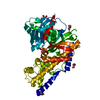
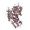
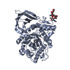
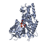
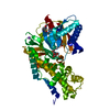
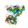
- PDB-4qs7: Arabidopsis Hexokinase 1 (AtHXK1) structure in glucose-bound form -
+ Open data

- Basic information
| Entry | Database: PDB / ID: 4qs7 | ||||||
|---|---|---|---|---|---|---|---|
| Title | Arabidopsis Hexokinase 1 (AtHXK1) structure in glucose-bound form | ||||||
 Components Components | Hexokinase-1 | ||||||
 Keywords Keywords | TRANSFERASE / Hexokinase / ATP-dependent / sugar sensor | ||||||
| Function / homology |  Function and homology information Function and homology informationhexose catabolic process / transpiration / stomatal closure / sugar mediated signaling pathway / hexokinase activity / regulation of secondary shoot formation / glucose mediated signaling pathway / hexokinase / fructokinase activity / glucokinase activity ...hexose catabolic process / transpiration / stomatal closure / sugar mediated signaling pathway / hexokinase activity / regulation of secondary shoot formation / glucose mediated signaling pathway / hexokinase / fructokinase activity / glucokinase activity / glucose 6-phosphate metabolic process / D-glucose binding / programmed cell death / plant-type vacuole / core promoter sequence-specific DNA binding / intracellular glucose homeostasis / glycolytic process / glucose metabolic process / mitochondrial outer membrane / regulation of transcription by RNA polymerase II / protein-containing complex / mitochondrion / zinc ion binding / ATP binding / nucleus / cytosol Similarity search - Function | ||||||
| Biological species |  | ||||||
| Method |  X-RAY DIFFRACTION / SYNCHROTRON / MOLECULAR REPLACEMENT / Resolution: 2.001 Å X-RAY DIFFRACTION / SYNCHROTRON / MOLECULAR REPLACEMENT / Resolution: 2.001 Å | ||||||
 Authors Authors | Feng, J. / Zhao, S. / Liu, L. | ||||||
 Citation Citation | Journal: Acta Crystallogr.,Sect.D / Year: 2015 Title: Biochemical and structural study of Arabidopsis hexokinase 1 Authors: Feng, J. / Zhao, S. / Chen, X. / Wang, W. / Dong, W. / Chen, J. / Shen, J.-R. / Liu, L. / Kuang, T. | ||||||
| History |
|
- Structure visualization
| Structure viewer | Molecule: MolmilJmol/JSmol |
|---|
- Downloads & links
-Download
| PDBx/mmCIF format | 4qs7.cif.gz | 193.4 KB | Display | PDBx/mmCIF format |
|---|---|---|---|---|
| PDB format | pdb4qs7.ent.gz | 151.9 KB | Display | PDB format |
| PDBx/mmJSON format | 4qs7.json.gz | Tree view | PDBx/mmJSON format | |
| Others |  Other downloads Other downloads |
-Validation report
| Arichive directory | https://data.pdbj.org/pub/pdb/validation_reports/qs/4qs7ftp://data.pdbj.org/pub/pdb/validation_reports/qs/4qs7 | HTTPS FTP |
|---|
-Related structure data
| Related structure data | 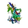 4qs8C 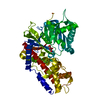 4qs9C 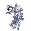 3o6wS C: citing same article ( S: Starting model for refinement |
|---|---|
| Similar structure data |


-Links
 PDBj
PDBj


- Assembly
| Deposited unit | 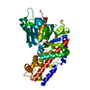
| ||||||||
|---|---|---|---|---|---|---|---|---|---|
| 1 |
| ||||||||
| Unit cell |
|
-Components
| #1: Protein | Mass: 51784.160 Da / Num. of mol.: 1 / Fragment: UNP residues 30-496 Source method: isolated from a genetically manipulated source Source: (gene. exp.)  |
|---|---|
| #2: Sugar | ChemComp-BGC /   Type: D-saccharide, beta linking / Mass: 180.156 Da / Num. of mol.: 1 Type: D-saccharide, beta linking / Mass: 180.156 Da / Num. of mol.: 1Source method: isolated from a genetically manipulated source Formula: C6H12O6 |
| #3: Water | ChemComp-HOH /  Mass: 18.015 Da / Num. of mol.: 287 / Source method: isolated from a natural source / Formula: H2O Mass: 18.015 Da / Num. of mol.: 287 / Source method: isolated from a natural source / Formula: H2O |
-Experimental details
-Experiment
| Experiment | Method: X-RAY DIFFRACTION / Number of used crystals: 1 |
|---|
- Sample preparation
| Crystal | Density Matthews: 1.83 Å3/Da / Density % sol: 32.93 % |
|---|---|
| Crystal grow | Temperature: 289 K / Method: vapor diffusion, hanging drop / pH: 7.5 Details: HEPES, 22% PEG 3350, 50uM glucose, pH 7.5, VAPOR DIFFUSION, HANGING DROP, temperature 289K |
-Data collection
| Diffraction | Mean temperature: 100 K |
|---|---|
| Diffraction source | Source: SYNCHROTRON / Site: SSRF  / Beamline: BL17U / Wavelength: 0.9793 Å / Beamline: BL17U / Wavelength: 0.9793 Å |
| Detector | Type: ADSC QUANTUM 315r / Detector: CCD / Date: May 20, 2013 |
| Radiation | Monochromator: double crystal / Protocol: SINGLE WAVELENGTH / Monochromatic (M) / Laue (L): M / Scattering type: x-ray |
| Radiation wavelength | Wavelength: 0.9793 Å / Relative weight: 1 |
| Reflection | Resolution: 2→50 Å / Num. all: 26533 / Num. obs: 26082 / % possible obs: 98.3 % / Observed criterion σ(F): 132739 / Observed criterion σ(I): 133077 / Biso Wilson estimate: 25.22 Å2 |
| Reflection shell | Resolution: 2→2.07 Å / % possible all: 91.6 |
- Processing
| Software |
| ||||||||||||||||||||||||||||||||||||||||||||||||||||||||||||||||||||||
|---|---|---|---|---|---|---|---|---|---|---|---|---|---|---|---|---|---|---|---|---|---|---|---|---|---|---|---|---|---|---|---|---|---|---|---|---|---|---|---|---|---|---|---|---|---|---|---|---|---|---|---|---|---|---|---|---|---|---|---|---|---|---|---|---|---|---|---|---|---|---|---|
| Refinement | Method to determine structure: MOLECULAR REPLACEMENT Starting model: 3o6w Resolution: 2.001→41.89 Å / FOM work R set: 0.8472 / SU ML: 0.21 / σ(F): 1.34 / Phase error: 21.68 / Stereochemistry target values: ML
| ||||||||||||||||||||||||||||||||||||||||||||||||||||||||||||||||||||||
| Solvent computation | Shrinkage radii: 0.9 Å / VDW probe radii: 1.11 Å / Solvent model: FLAT BULK SOLVENT MODEL | ||||||||||||||||||||||||||||||||||||||||||||||||||||||||||||||||||||||
| Displacement parameters | Biso max: 86.43 Å2 / Biso mean: 26.66 Å2 / Biso min: 11.01 Å2 | ||||||||||||||||||||||||||||||||||||||||||||||||||||||||||||||||||||||
| Refinement step | Cycle: LAST / Resolution: 2.001→41.89 Å
| ||||||||||||||||||||||||||||||||||||||||||||||||||||||||||||||||||||||
| Refine LS restraints |
| ||||||||||||||||||||||||||||||||||||||||||||||||||||||||||||||||||||||
| LS refinement shell | Refine-ID: X-RAY DIFFRACTION / Total num. of bins used: 9
| ||||||||||||||||||||||||||||||||||||||||||||||||||||||||||||||||||||||
| Refinement TLS params. | Method: refined / Origin x: 10.4723 Å / Origin y: 7.0838 Å / Origin z: 8.4843 Å
| ||||||||||||||||||||||||||||||||||||||||||||||||||||||||||||||||||||||
| Refinement TLS group | Selection details: ALL |
