- PDB-4qhz: Crystal structure of a putative glycosyl hydrolase (BDI_3914) fro... -
+
Open data
ID or keywords:
Loading...
-
Basic information
Entry
Database: PDB / ID: 4qhz
Title
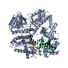
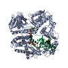
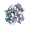
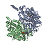
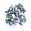
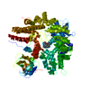
Crystal structure of a putative glycosyl hydrolase (BDI_3914) from Parabacteroides distasonis ATCC 8503 at 2.13 A resolution
Components
putative glycosyl hydrolase
Keywords
HYDROLASE / PF06439 family protein / DUF1080 / Structural Genomics / Joint Center for Structural Genomics / JCSG / Protein Structure Initiative / PSI-BIOLOGY
Mass: 18.015 Da / Num. of mol.: 910 / Source method: isolated from a natural source / Formula: H2O
Sequence details
1.THIS CONSTRUCT WAS EXPRESSED WITH AN N-TERMINAL PURIFICATION TAG MGSDKIHHHHHHENLYFQG. THE TAG WAS ...1.THIS CONSTRUCT WAS EXPRESSED WITH AN N-TERMINAL PURIFICATION TAG MGSDKIHHHHHHENLYFQG. THE TAG WAS REMOVED WITH TEV PROTEASE LEAVING ONLY A GLYCINE (0) FOLLOWED BY RESIDUES 21-265 OF THE TARGET SEQUENCE. 2. THE PROTEIN WAS REDUCTIVELY METHYLATED PRIOR TO CRYSTALLIZATION. ALL LYSINE RESIDUES (EXCEPT RESIDUES 102, 169, AND 197) HAVE BEEN MODELED AS MLY.
-
Experimental details
-
Experiment
Experiment
Method: X-RAY DIFFRACTION / Number of used crystals: 2
-
Sample preparation
Crystal
Density Matthews: 2.5 Å3/Da / Density % sol: 50.84 % Description: THE STRUCTURE WAS SOLVED BY THREE WAVELENGTH MAD METHOD AT 2.40A RESOLUTION. THE STRUCTURE WAS REFINED AT 2.13A RESOLUTION AGAINST A DATASET COLLECTED FROM A DIFFERENT CRYSTAL.
Resolution: 2.13→48.254 Å / Num. obs: 58318 / % possible obs: 95 % / Observed criterion σ(I): -3 / Biso Wilson estimate: 26.92 Å2 / Rmerge(I) obs: 0.111 / Net I/σ(I): 6.96
Reflection shell
Diffraction-ID: 1,2
Resolution (Å)
Highest resolution (Å)
Rmerge(I) obs
Mean I/σ(I) obs
Num. measured obs
Num. unique obs
% possible all
2.13-2.21
0.477
2.1
14623
6159
96.6
2.21-2.29
0.432
2.4
12372
5278
94.2
2.29-2.4
0.355
2.9
14412
6130
95.4
2.4-2.52
0.274
3.6
13800
5679
97.2
2.52-2.68
0.229
4.2
14279
5960
96
2.68-2.89
0.168
5.5
13526
5743
92.7
2.89-3.18
0.119
7.7
14551
5943
96.7
3.18-3.64
0.072
11.4
13932
5769
94.2
3.64-4.57
0.053
14.7
14198
5775
94.8
4.57
0.056
15.1
14117
5820
92.4
-
Phasing
Phasing
Method: MAD
-
Processing
Software
Name
Version
Classification
NB
MolProbity
3beta29
modelbuilding
PDB_EXTRACT
3.1
dataextraction
SOLVE
phasing
REFMAC
5.7.0032
refinement
XDS
datareduction
XSCALE
datascaling
Refinement
Method to determine structure: MAD / Resolution: 2.13→48.254 Å / Cor.coef. Fo:Fc: 0.954 / Cor.coef. Fo:Fc free: 0.939 / Occupancy max: 1 / Occupancy min: 0.4 / SU B: 8.623 / SU ML: 0.12 / Cross valid method: THROUGHOUT / σ(F): 0 / ESU R: 0.237 / ESU R Free: 0.176 Stereochemistry target values: MAXIMUM LIKELIHOOD WITH PHASES Details: 1. HYDROGENS HAVE BEEN ADDED IN THE RIDING POSITIONS. 2. ATOM RECORD CONTAINS SUM OF TLS AND RESIDUAL B FACTORS. ANISOU RECORD CONTAINS SUM OF TLS AND RESIDUAL U FACTORS. 3. WATERS WERE ...Details: 1. HYDROGENS HAVE BEEN ADDED IN THE RIDING POSITIONS. 2. ATOM RECORD CONTAINS SUM OF TLS AND RESIDUAL B FACTORS. ANISOU RECORD CONTAINS SUM OF TLS AND RESIDUAL U FACTORS. 3. WATERS WERE EXCLUDED FROM AUTOMATIC TLS ASSIGNMENT. 4. A MET-INHIBITION PROTOCOL WAS USED FOR SELENOMETHIONINE INCORPORATION DURING PROTEIN EXPRESSION. THE OCCUPANCY OF THE SE ATOMS IN THE MSE RESIDUES WAS REDUCED TO 0.75 FOR THE REDUCED SCATTERING POWER DUE TO PARTIAL S-MET INCORPORATION. 5. THE PROTEIN WAS REDUCTIVELY METHYLATED PRIOR RO CRYSTALLIZATION. THEREFORE ALL LYSINE RESIDUES (EXCEPT RESIDUES 102, 169, AND 197) HAVE BEEN MODELED AS MLY. ELECTRON DENSITY SUGGESTS THAT THESE THREE RESIDUES ARE UNMODIFIED. 6. MAGNESIUM (MG) IONS AND 1,2-ETHANEDIOL (EDO) MOLECULES FROM THE CRYSTALLIZATION/CRYOPROTECTION SOLUTION ARE MODELED. 7. THE REFINEMENT WAS RESTRAINED AGAINST THE MAD PHASES FROM A CRYSTAL THAT WAS USED TO OBTAIN THE STRUCTURE SOLUTION.
Rfactor
Num. reflection
% reflection
Selection details
Rfree
0.2035
2948
5.1 %
RANDOM
Rwork
0.1707
-
-
-
obs
0.1724
55339
95.03 %
-
Solvent computation
Ion probe radii: 0.8 Å / Shrinkage radii: 0.8 Å / VDW probe radii: 1.2 Å / Solvent model: BABINET MODEL WITH MASK
In the structure databanks used in Yorodumi, some data are registered as the other names, "COVID-19 virus" and "2019-nCoV". Here are the details of the virus and the list of structure data.
Jan 31, 2019. EMDB accession codes are about to change! (news from PDBe EMDB page)
EMDB accession codes are about to change! (news from PDBe EMDB page)
The allocation of 4 digits for EMDB accession codes will soon come to an end. Whilst these codes will remain in use, new EMDB accession codes will include an additional digit and will expand incrementally as the available range of codes is exhausted. The current 4-digit format prefixed with “EMD-” (i.e. EMD-XXXX) will advance to a 5-digit format (i.e. EMD-XXXXX), and so on. It is currently estimated that the 4-digit codes will be depleted around Spring 2019, at which point the 5-digit format will come into force.
The EM Navigator/Yorodumi systems omit the EMD- prefix.
Related info.:Q: What is EMD? / ID/Accession-code notation in Yorodumi/EM Navigator
Yorodumi is a browser for structure data from EMDB, PDB, SASBDB, etc.
This page is also the successor to EM Navigator detail page, and also detail information page/front-end page for Omokage search.
The word "yorodu" (or yorozu) is an old Japanese word meaning "ten thousand". "mi" (miru) is to see.
Related info.:EMDB / PDB / SASBDB / Comparison of 3 databanks / Yorodumi Search / Aug 31, 2016. New EM Navigator & Yorodumi / Yorodumi Papers / Jmol/JSmol / Function and homology information / Changes in new EM Navigator and Yorodumi
 Movie
Movie Controller

 Yorodumi
 Open data
 Basic information
 Components
 Keywords
 Function and homology information
 Parabacteroides distasonis (bacteria)
 X-RAY DIFFRACTION /
  Authors
 Citation
 Structure visualization
 Downloads & links
 Other downloads
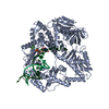


 PDBj
PDBj


 Assembly
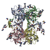

 Mass: 62.068 Da / Num. of mol.: 3 / Source method: obtained synthetically / Formula: C2H6O2
 Mass: 24.305 Da / Num. of mol.: 4 / Source method: obtained synthetically / Formula: Mg
 Mass: 18.015 Da / Num. of mol.: 910 / Source method: isolated from a natural source / Formula: H2O
 Sample preparation
 Processing