 Movie
Movie Controller

[English] 日本語
 Yorodumi
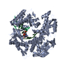
- PDB-3k57: Crystal structure of E.coli Pol II-normal DNA-dATP ternary complex -
+ Open data

- Basic information
| Entry | Database: PDB / ID: 3k57 | ||||||
|---|---|---|---|---|---|---|---|
| Title | Crystal structure of E.coli Pol II-normal DNA-dATP ternary complex | ||||||
 Components Components |
| ||||||
 Keywords Keywords | TRANSFERASE/DNA / PROTEIN-DNA COMPLEX / DNA damage / DNA repair / DNA-binding / DNA-directed DNA polymerase / Nucleotidyltransferase / SOS response / Transferase / TRANSFERASE-DNA COMPLEX | ||||||
| Function / homology |  Function and homology information Function and homology informationDNA replication proofreading / 3'-5'-DNA exonuclease activity / SOS response / DNA-templated DNA replication / DNA-directed DNA polymerase / DNA-directed DNA polymerase activity / nucleotide binding / DNA binding Similarity search - Function | ||||||
| Biological species |  | ||||||
| Method |  X-RAY DIFFRACTION / MOLECULAR REPLACEMENT / Resolution: 2.08 Å X-RAY DIFFRACTION / MOLECULAR REPLACEMENT / Resolution: 2.08 Å | ||||||
 Authors Authors | Yang, W. / Wang, F. | ||||||
 Citation Citation | Journal: Cell(Cambridge,Mass.) / Year: 2009 Title: Structural insight into translesion synthesis by DNA Pol II Authors: Wang, F. / Yang, W. | ||||||
| History |
|
- Structure visualization
| Structure viewer | Molecule: MolmilJmol/JSmol |
|---|
- Downloads & links
-Download
| PDBx/mmCIF format | 3k57.cif.gz | 203.6 KB | Display | PDBx/mmCIF format |
|---|---|---|---|---|
| PDB format | pdb3k57.ent.gz | 153.9 KB | Display | PDB format |
| PDBx/mmJSON format | 3k57.json.gz | Tree view | PDBx/mmJSON format | |
| Others |  Other downloads Other downloads |
-Validation report
| Arichive directory | https://data.pdbj.org/pub/pdb/validation_reports/k5/3k57ftp://data.pdbj.org/pub/pdb/validation_reports/k5/3k57 | HTTPS FTP |
|---|
-Related structure data
| Related structure data |  3k58C 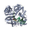 3k59C  3k5lC 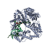 3k5mC  3k5nC 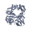 3k5oC  3maqC 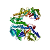 1q8iS C: citing same article ( S: Starting model for refinement |
|---|---|
| Similar structure data |


-Links
 PDBj
PDBj


- Assembly
| Deposited unit | 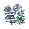
| ||||||||
|---|---|---|---|---|---|---|---|---|---|
| 1 |
| ||||||||
| Unit cell |
|
-Components
-Protein , 1 types, 1 molecules A
| #1: Protein | Mass: 90459.734 Da / Num. of mol.: 1 / Mutation: D335N Source method: isolated from a genetically manipulated source Source: (gene. exp.) Plasmid: Modified PET28 with prescission protease cleavage site Production host: |
|---|
-DNA chain , 2 types, 2 molecules TP
| #2: DNA chain | Mass: 5556.603 Da / Num. of mol.: 1 / Source method: obtained synthetically / Details: Chemical synthesis |
|---|---|
| #3: DNA chain | Mass: 3951.586 Da / Num. of mol.: 1 / Source method: obtained synthetically / Details: Chemical synthesis |
-Non-polymers , 3 types, 528 molecules 


| #4: Chemical | ChemComp-DTP /  Mass: 491.182 Da / Num. of mol.: 1 / Source method: obtained synthetically / Formula: C10H16N5O12P3 Mass: 491.182 Da / Num. of mol.: 1 / Source method: obtained synthetically / Formula: C10H16N5O12P3 | ||
|---|---|---|---|
| #5: Chemical |  Mass: 24.305 Da / Num. of mol.: 2 / Source method: obtained synthetically / Formula: Mg Mass: 24.305 Da / Num. of mol.: 2 / Source method: obtained synthetically / Formula: Mg#6: Water | ChemComp-HOH / | Mass: 18.015 Da / Num. of mol.: 525 / Source method: isolated from a natural source / Formula: H2O |
-Experimental details
-Experiment
| Experiment | Method: X-RAY DIFFRACTION / Number of used crystals: 1 |
|---|
- Sample preparation
| Crystal | Density Matthews: 2.53 Å3/Da / Density % sol: 51.31 % |
|---|---|
| Crystal grow | Temperature: 293 K / Method: vapor diffusion, hanging drop / pH: 6.5 Details: 100 mM MES, 20% PEGmme5000, 0.2 M ammonium sulfate, pH 6.5, VAPOR DIFFUSION, HANGING DROP, temperature 293K |
-Data collection
| Diffraction | Mean temperature: 95 K |
|---|---|
| Diffraction source | Source: ROTATING ANODE / Type: RIGAKU / Wavelength: 1.5418 Å |
| Detector | Type: RIGAKU RAXIS IV / Detector: IMAGE PLATE / Date: Oct 12, 2006 |
| Radiation | Protocol: SINGLE WAVELENGTH / Monochromatic (M) / Laue (L): M / Scattering type: x-ray |
| Radiation wavelength | Wavelength: 1.5418 Å / Relative weight: 1 |
| Reflection | Resolution: 2.08→20 Å / Num. all: 61590 / Num. obs: 57617 / % possible obs: 93.5 % / Observed criterion σ(I): -3 / Redundancy: 2.4 % / Rmerge(I) obs: 0.07 / Net I/σ(I): 13.6 |
| Reflection shell | Resolution: 2.08→2.15 Å / Redundancy: 1.9 % / Rmerge(I) obs: 0.478 / Mean I/σ(I) obs: 1.8 / Num. unique all: 6065 / % possible all: 87 |
- Processing
| Software |
| ||||||||||||||||||||
|---|---|---|---|---|---|---|---|---|---|---|---|---|---|---|---|---|---|---|---|---|---|
| Refinement | Method to determine structure: MOLECULAR REPLACEMENT Starting model: PDB ENTRY 1Q8I Resolution: 2.08→20 Å / Cross valid method: THROUGHOUT / σ(F): 0 / Stereochemistry target values: Engh & Huber
| ||||||||||||||||||||
| Refine analyze |
| ||||||||||||||||||||
| Refinement step | Cycle: LAST / Resolution: 2.08→20 Å
| ||||||||||||||||||||
| Refine LS restraints |
| ||||||||||||||||||||
| LS refinement shell | Resolution: 2.08→2.15 Å
|
