 Movie
Movie Controller


+ Open data

- Basic information
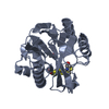
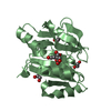
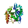
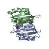
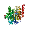
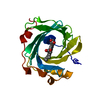
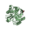
| Entry | Database: PDB / ID: 4qc6 | ||||||
|---|---|---|---|---|---|---|---|
| Title | Crystal structure of aminoglycoside 6'-acetyltransferase-Ie | ||||||
 Components Components | Bifunctional AAC/APH | ||||||
 Keywords Keywords | transferase/antibiotic / antibiotic resistance / GNAT family / acetyltransferase / acetylcoenzyme-A / aminoglycoside / transferase / transferase-antibiotic complex | ||||||
| Function / homology |  Function and homology information Function and homology informationaminoglycoside 2''-phosphotransferase / aminoglycoside phosphotransferase activity / : / Transferases; Acyltransferases; Transferring groups other than aminoacyl groups / response to antibiotic / ATP binding / cytoplasm Similarity search - Function | ||||||
| Biological species |  Staphylococcus warneri (bacteria) Staphylococcus warneri (bacteria) | ||||||
| Method |  X-RAY DIFFRACTION / SYNCHROTRON / MOLECULAR REPLACEMENT / Resolution: 1.3 Å X-RAY DIFFRACTION / SYNCHROTRON / MOLECULAR REPLACEMENT / Resolution: 1.3 Å | ||||||
 Authors Authors | Smith, C.A. / Toth, M. / Weiss, T.M. / Frase, H. / Vakulenko, S.B. | ||||||
 Citation Citation | Journal: Acta Crystallogr.,Sect.D / Year: 2014 Title: Structure of the bifunctional aminoglycoside-resistance enzyme AAC(6')-Ie-APH(2'')-Ia revealed by crystallographic and small-angle X-ray scattering analysis. Authors: Smith, C.A. / Toth, M. / Weiss, T.M. / Frase, H. / Vakulenko, S.B. | ||||||
| History |
|
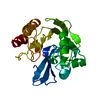
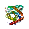
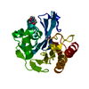
- Structure visualization
| Structure viewer | Molecule: MolmilJmol/JSmol |
|---|
- Downloads & links
-Download
| PDBx/mmCIF format | 4qc6.cif.gz | 195.6 KB | Display | PDBx/mmCIF format |
|---|---|---|---|---|
| PDB format | pdb4qc6.ent.gz | 156.9 KB | Display | PDB format |
| PDBx/mmJSON format | 4qc6.json.gz | Tree view | PDBx/mmJSON format | |
| Others |  Other downloads Other downloads |
-Validation report
| Arichive directory | https://data.pdbj.org/pub/pdb/validation_reports/qc/4qc6ftp://data.pdbj.org/pub/pdb/validation_reports/qc/4qc6 | HTTPS FTP |
|---|
-Related structure data
| Similar structure data |
|---|
-Links
 PDBj
PDBj

- Assembly
| Deposited unit | 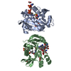
| ||||||||
|---|---|---|---|---|---|---|---|---|---|
| 1 | 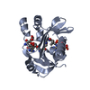
| ||||||||
| 2 | 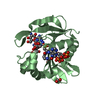
| ||||||||
| Unit cell |
|
-Components
| #1: Protein | Mass: 21735.715 Da / Num. of mol.: 2 Source method: isolated from a genetically manipulated source Source: (gene. exp.) Staphylococcus warneri (bacteria) / Gene: aacA-aphD / Production host: References: UniProt: Q7ATH7, Transferases; Acyltransferases; Transferring groups other than aminoacyl groups, Transferases; Transferring phosphorus-containing groups; Phosphotransferases with an alcohol group as acceptor #2: Chemical |   Mass: 484.499 Da / Num. of mol.: 2 / Source method: obtained synthetically / Formula: C18H36N4O11 / Comment: antibiotic*YM Mass: 484.499 Da / Num. of mol.: 2 / Source method: obtained synthetically / Formula: C18H36N4O11 / Comment: antibiotic*YM#3: Chemical |   Mass: 799.533 Da / Num. of mol.: 2 / Source method: obtained synthetically / Formula: C21H36N7O18P3S Mass: 799.533 Da / Num. of mol.: 2 / Source method: obtained synthetically / Formula: C21H36N7O18P3S#4: Chemical | ChemComp-FMT /   Mass: 46.025 Da / Num. of mol.: 5 / Source method: obtained synthetically / Formula: CH2O2 Mass: 46.025 Da / Num. of mol.: 5 / Source method: obtained synthetically / Formula: CH2O2#5: Water | ChemComp-HOH / |  Mass: 18.015 Da / Num. of mol.: 705 / Source method: isolated from a natural source / Formula: H2O Mass: 18.015 Da / Num. of mol.: 705 / Source method: isolated from a natural source / Formula: H2O |
|---|
-Experimental details
-Experiment
| Experiment | Method: X-RAY DIFFRACTION / Number of used crystals: 1 |
|---|
- Sample preparation
| Crystal | Density Matthews: 2.64 Å3/Da / Density % sol: 53.46 % |
|---|---|
| Crystal grow | Temperature: 278 K / Method: vapor diffusion, sitting drop Details: unbuffered 0.2 M ammonium formate, 20% PEG 3350, VAPOR DIFFUSION, SITTING DROP, temperature 278K |
-Data collection
| Diffraction | Mean temperature: 100 K |
|---|---|
| Diffraction source | Source: SYNCHROTRON / Site: SSRL  / Beamline: BL12-2 / Wavelength: 0.9795 Å / Beamline: BL12-2 / Wavelength: 0.9795 Å |
| Detector | Type: DECTRIS PILATUS 6M / Detector: PIXEL / Date: Dec 9, 2011 |
| Radiation | Monochromator: Si(111) / Protocol: SINGLE WAVELENGTH / Monochromatic (M) / Laue (L): M / Scattering type: x-ray |
| Radiation wavelength | Wavelength: 0.9795 Å / Relative weight: 1 |
| Reflection | Resolution: 1.3→19.941 Å / Num. all: 112448 / Num. obs: 112448 / % possible obs: 98.7 % / Observed criterion σ(F): 0 / Observed criterion σ(I): 0 / Redundancy: 5.3 % / Biso Wilson estimate: 14.6 Å2 / Rmerge(I) obs: 0.064 / Net I/σ(I): 13.1 |
| Reflection shell | Resolution: 1.3→1.35 Å / Rmerge(I) obs: 0.371 / Mean I/σ(I) obs: 2.4 / % possible all: 87.2 |
- Processing
| Software |
| |||||||||||||||||||||||||||||||||||||||||||||||||||||||||||||||||||||||||||||||||||||||||||||||||||||||||||||||||||||||||||||||||||||||||||||||||||||||||||||||||||||||||||||||||||||||||||||||||||||||||||||||||||||||||
|---|---|---|---|---|---|---|---|---|---|---|---|---|---|---|---|---|---|---|---|---|---|---|---|---|---|---|---|---|---|---|---|---|---|---|---|---|---|---|---|---|---|---|---|---|---|---|---|---|---|---|---|---|---|---|---|---|---|---|---|---|---|---|---|---|---|---|---|---|---|---|---|---|---|---|---|---|---|---|---|---|---|---|---|---|---|---|---|---|---|---|---|---|---|---|---|---|---|---|---|---|---|---|---|---|---|---|---|---|---|---|---|---|---|---|---|---|---|---|---|---|---|---|---|---|---|---|---|---|---|---|---|---|---|---|---|---|---|---|---|---|---|---|---|---|---|---|---|---|---|---|---|---|---|---|---|---|---|---|---|---|---|---|---|---|---|---|---|---|---|---|---|---|---|---|---|---|---|---|---|---|---|---|---|---|---|---|---|---|---|---|---|---|---|---|---|---|---|---|---|---|---|---|---|---|---|---|---|---|---|---|---|---|---|---|---|---|---|---|
| Refinement | Method to determine structure: MOLECULAR REPLACEMENT / Resolution: 1.3→19.941 Å / SU ML: 0.09 / σ(F): 1.37 / Phase error: 14.85 / Stereochemistry target values: ML
| |||||||||||||||||||||||||||||||||||||||||||||||||||||||||||||||||||||||||||||||||||||||||||||||||||||||||||||||||||||||||||||||||||||||||||||||||||||||||||||||||||||||||||||||||||||||||||||||||||||||||||||||||||||||||
| Solvent computation | Shrinkage radii: 0.9 Å / VDW probe radii: 1.11 Å / Solvent model: FLAT BULK SOLVENT MODEL | |||||||||||||||||||||||||||||||||||||||||||||||||||||||||||||||||||||||||||||||||||||||||||||||||||||||||||||||||||||||||||||||||||||||||||||||||||||||||||||||||||||||||||||||||||||||||||||||||||||||||||||||||||||||||
| Refinement step | Cycle: LAST / Resolution: 1.3→19.941 Å
| |||||||||||||||||||||||||||||||||||||||||||||||||||||||||||||||||||||||||||||||||||||||||||||||||||||||||||||||||||||||||||||||||||||||||||||||||||||||||||||||||||||||||||||||||||||||||||||||||||||||||||||||||||||||||
| Refine LS restraints |
| |||||||||||||||||||||||||||||||||||||||||||||||||||||||||||||||||||||||||||||||||||||||||||||||||||||||||||||||||||||||||||||||||||||||||||||||||||||||||||||||||||||||||||||||||||||||||||||||||||||||||||||||||||||||||
| LS refinement shell |
|
