 Movie
Movie Controller

[English] 日本語
 Yorodumi
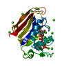
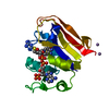
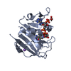
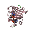
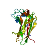
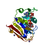
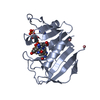
- PDB-4q67: Staphylococcus aureus F98Y mutant dihydrofolate reductase complex... -
+ Open data

- Basic information
| Entry | Database: PDB / ID: 4q67 | ||||||
|---|---|---|---|---|---|---|---|
| Title | Staphylococcus aureus F98Y mutant dihydrofolate reductase complexed with NADPH | ||||||
 Components Components | Dihydrofolate reductase | ||||||
 Keywords Keywords | OXIDOREDUCTASE | ||||||
| Function / homology | Dihydrofolate Reductase, subunit A / Dihydrofolate Reductase, subunit A / 3-Layer(aba) Sandwich / Alpha Beta / NADP NICOTINAMIDE-ADENINE-DINUCLEOTIDE PHOSPHATE / :  Function and homology information Function and homology information | ||||||
| Biological species |  Staphylococcus aureus MUF168 (bacteria) Staphylococcus aureus MUF168 (bacteria) | ||||||
| Method |  X-RAY DIFFRACTION / MOLECULAR REPLACEMENT / Resolution: 2.04 Å X-RAY DIFFRACTION / MOLECULAR REPLACEMENT / Resolution: 2.04 Å | ||||||
 Authors Authors | Reeve, S.M. / Anderson, A.C. | ||||||
 Citation Citation | Journal: Proc.Natl.Acad.Sci.USA / Year: 2015 Title: Protein design algorithms predict viable resistance to an experimental antifolate. Authors: Reeve, S.M. / Gainza, P. / Frey, K.M. / Georgiev, I. / Donald, B.R. / Anderson, A.C. | ||||||
| History |
|
- Structure visualization
| Structure viewer | Molecule: MolmilJmol/JSmol |
|---|
- Downloads & links
-Download
| PDBx/mmCIF format | 4q67.cif.gz | 85.4 KB | Display | PDBx/mmCIF format |
|---|---|---|---|---|
| PDB format | pdb4q67.ent.gz | 63.9 KB | Display | PDB format |
| PDBx/mmJSON format | 4q67.json.gz | Tree view | PDBx/mmJSON format | |
| Others |  Other downloads Other downloads |
-Validation report
| Summary document | 4q67_validation.pdf.gz | 718.3 KB | Display | wwPDB validaton report |
|---|---|---|---|---|
| Full document | 4q67_full_validation.pdf.gz | 718.3 KB | Display | |
| Data in XML | 4q67_validation.xml.gz | 9.1 KB | Display | |
| Data in CIF | 4q67_validation.cif.gz | 12.2 KB | Display | |
| Arichive directory | https://data.pdbj.org/pub/pdb/validation_reports/q6/4q67ftp://data.pdbj.org/pub/pdb/validation_reports/q6/4q67 | HTTPS FTP |
-Related structure data
| Related structure data | 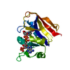 4q6aC 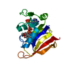 3fqoS C: citing same article ( S: Starting model for refinement |
|---|---|
| Similar structure data |


-Links
 PDBj
PDBj- Assembly
| Deposited unit | 
| ||||||||
|---|---|---|---|---|---|---|---|---|---|
| 1 |
| ||||||||
| Unit cell |
|
-Components
| #1: Protein | Mass: 18405.027 Da / Num. of mol.: 1 / Mutation: F98Y Source method: isolated from a genetically manipulated source Source: (gene. exp.) Staphylococcus aureus MUF168 (bacteria)Strain: ATCC 43300 / Gene: dfrB, SaDHFR, Y000_11620 / Plasmid: pET-41a(+) / Production host: |
|---|---|
| #2: Chemical | ChemComp-NAP /   Mass: 743.405 Da / Num. of mol.: 1 / Source method: obtained synthetically / Formula: C21H28N7O17P3 Mass: 743.405 Da / Num. of mol.: 1 / Source method: obtained synthetically / Formula: C21H28N7O17P3 |
| #3: Water | ChemComp-HOH /  Mass: 18.015 Da / Num. of mol.: 103 / Source method: isolated from a natural source / Formula: H2O Mass: 18.015 Da / Num. of mol.: 103 / Source method: isolated from a natural source / Formula: H2O |
-Experimental details
-Experiment
| Experiment | Method: X-RAY DIFFRACTION / Number of used crystals: 1 |
|---|
- Sample preparation
| Crystal | Density Matthews: 2.65 Å3/Da / Density % sol: 53.52 % |
|---|---|
| Crystal grow | Temperature: 277 K / Method: vapor diffusion, hanging drop / pH: 6 Details: 0.1M Sodium Acetate, 0.1M MES, 0.15%, 13% PEG 10,000, gamma-butyrolacetone, pH 6.0, VAPOR DIFFUSION, HANGING DROP, temperature 277K |
-Data collection
| Diffraction | Mean temperature: 100 K |
|---|---|
| Diffraction source | Source: ROTATING ANODE / Type: RIGAKU MICROMAX-007 HF / Wavelength: 1.54178 Å |
| Detector | Type: RIGAKU RAXIS IV / Detector: IMAGE PLATE / Date: Jan 11, 2013 / Details: Varimax optics |
| Radiation | Monochromator: Copper Anode / Protocol: SINGLE WAVELENGTH / Monochromatic (M) / Laue (L): M / Scattering type: x-ray |
| Radiation wavelength | Wavelength: 1.54178 Å / Relative weight: 1 |
| Reflection | Resolution: 2.04→42.3 Å / Num. all: 13109 / Num. obs: 12466 / % possible obs: 99 % / Observed criterion σ(F): 0 / Observed criterion σ(I): 0 / Redundancy: 10.22 % / Biso Wilson estimate: 26.05 Å2 / Rmerge(I) obs: 0.07 / Net I/σ(I): 16.9 |
| Reflection shell | Resolution: 2.04→2.11 Å / Redundancy: 1044 % / Rmerge(I) obs: 0.34 / Mean I/σ(I) obs: 5.6 / Num. unique all: 1286 / % possible all: 98.8 |
- Processing
| Software |
| ||||||||||||||||||||||||||||||||||||||||||
|---|---|---|---|---|---|---|---|---|---|---|---|---|---|---|---|---|---|---|---|---|---|---|---|---|---|---|---|---|---|---|---|---|---|---|---|---|---|---|---|---|---|---|---|
| Refinement | Method to determine structure: MOLECULAR REPLACEMENT Starting model: pdb entry 3FQO Resolution: 2.04→37.18 Å / SU ML: 0.18 / σ(F): 1.34 / σ(I): 2.1 / Phase error: 22.09 / Stereochemistry target values: ML
| ||||||||||||||||||||||||||||||||||||||||||
| Solvent computation | Shrinkage radii: 0.9 Å / VDW probe radii: 1.11 Å / Solvent model: FLAT BULK SOLVENT MODEL | ||||||||||||||||||||||||||||||||||||||||||
| Refinement step | Cycle: LAST / Resolution: 2.04→37.18 Å
| ||||||||||||||||||||||||||||||||||||||||||
| Refine LS restraints |
| ||||||||||||||||||||||||||||||||||||||||||
| LS refinement shell | Refine-ID: X-RAY DIFFRACTION
| ||||||||||||||||||||||||||||||||||||||||||
| Refinement TLS params. | Method: refined / Origin x: 1.7573 Å / Origin y: 27.4759 Å / Origin z: 3.2776 Å
| ||||||||||||||||||||||||||||||||||||||||||
| Refinement TLS group | Selection details: all |
