 Movie
Movie Controller

[English] 日本語
 Yorodumi
- PDB-4q5y: Crystal structure of extended-Tudor 10-11 of Drosophila melanogaster -
+ Open data

- Basic information
| Entry | Database: PDB / ID: 4q5y | ||||||
|---|---|---|---|---|---|---|---|
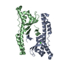
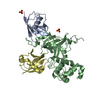
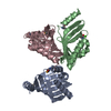
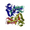
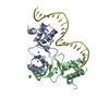
| Title | Crystal structure of extended-Tudor 10-11 of Drosophila melanogaster | ||||||
 Components Components | Maternal protein tudor | ||||||
 Keywords Keywords | TRANSCRIPTION / tudor domain / recognize sDMA of Aubergine / sDMA of Aubergine / nucleus | ||||||
| Function / homology |  Function and homology information Function and homology informationmitochondrial RNA localization / pole cell development / positive regulation of post-transcriptional gene silencing by RNA / P granule assembly / pole plasm / methylation-dependent protein binding / pole plasm assembly / pole cell formation / P granule organization / secondary piRNA processing ...mitochondrial RNA localization / pole cell development / positive regulation of post-transcriptional gene silencing by RNA / P granule assembly / pole plasm / methylation-dependent protein binding / pole plasm assembly / pole cell formation / P granule organization / secondary piRNA processing / intracellular mRNA localization / P granule / oogenesis / germ cell development / perinuclear region of cytoplasm / mitochondrion / nucleus / cytoplasm / cytosol Similarity search - Function | ||||||
| Biological species |  | ||||||
| Method |  X-RAY DIFFRACTION / SYNCHROTRON / MOLECULAR REPLACEMENT / Resolution: 3 Å X-RAY DIFFRACTION / SYNCHROTRON / MOLECULAR REPLACEMENT / Resolution: 3 Å | ||||||
 Authors Authors | Liu, H. / Ren, R. / Wang, W. / Wang, M. / Yang, N. / Dong, Y. / Gong, W. / Lehmann, R. / Xu, R.M. | ||||||
 Citation Citation | Journal: Cell Res. / Year: 2014 Title: Structure and domain organization of Drosophila Tudor Authors: Ren, R. / Liu, H. / Wang, W. / Wang, M. / Yang, N. / Dong, Y.H. / Gong, W. / Lehmann, R. / Xu, R.M. | ||||||
| History |
|
- Structure visualization
| Structure viewer | Molecule: MolmilJmol/JSmol |
|---|
- Downloads & links
-Download
| PDBx/mmCIF format | 4q5y.cif.gz | 82.2 KB | Display | PDBx/mmCIF format |
|---|---|---|---|---|
| PDB format | pdb4q5y.ent.gz | 60.1 KB | Display | PDB format |
| PDBx/mmJSON format | 4q5y.json.gz | Tree view | PDBx/mmJSON format | |
| Others |  Other downloads Other downloads |
-Validation report
| Arichive directory | https://data.pdbj.org/pub/pdb/validation_reports/q5/4q5yftp://data.pdbj.org/pub/pdb/validation_reports/q5/4q5y | HTTPS FTP |
|---|
-Related structure data
| Related structure data |  4q5wC  3ntkS C: citing same article ( S: Starting model for refinement |
|---|---|
| Similar structure data |


-Links
 PDBj
PDBj- Assembly
| Deposited unit | 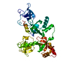
| ||||||||
|---|---|---|---|---|---|---|---|---|---|
| 1 |
| ||||||||
| 2 | 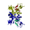
| ||||||||
| Unit cell |
|
-Components
| #1: Protein | Mass: 40323.484 Da / Num. of mol.: 1 / Fragment: extended-Tudor 10-11, UNP residues 2164-2515 Source method: isolated from a genetically manipulated source Source: (gene. exp.)  |
|---|---|
| #2: Water | ChemComp-HOH /  Mass: 18.015 Da / Num. of mol.: 56 / Source method: isolated from a natural source / Formula: H2O Mass: 18.015 Da / Num. of mol.: 56 / Source method: isolated from a natural source / Formula: H2O |
| Has protein modification | Y |
-Experimental details
-Experiment
| Experiment | Method: X-RAY DIFFRACTION / Number of used crystals: 1 |
|---|
- Sample preparation
| Crystal | Density Matthews: 4.2 Å3/Da / Density % sol: 70.74 % |
|---|---|
| Crystal grow | Temperature: 289 K / Method: vapor diffusion, hanging drop / pH: 7 Details: 60% Tacsimate, pH 7.0, VAPOR DIFFUSION, HANGING DROP, temperature 289K |
-Data collection
| Diffraction | Mean temperature: 100 K |
|---|---|
| Diffraction source | Source: SYNCHROTRON / Site: SSRF  / Beamline: BL17U / Wavelength: 0.99985 Å / Beamline: BL17U / Wavelength: 0.99985 Å |
| Detector | Type: RAYONIX MX-225 / Detector: CCD / Date: Nov 10, 2009 / Details: mirrors |
| Radiation | Monochromator: double crystal / Protocol: SINGLE WAVELENGTH / Monochromatic (M) / Laue (L): M / Scattering type: x-ray |
| Radiation wavelength | Wavelength: 0.99985 Å / Relative weight: 1 |
| Reflection | Resolution: 3→50 Å / Num. all: 13994 / Num. obs: 13606 / % possible obs: 97.2 % / Observed criterion σ(F): 2 / Observed criterion σ(I): 2 / Redundancy: 5.1 % / Rmerge(I) obs: 0.068 / Net I/σ(I): 21.1 |
| Reflection shell | Resolution: 3→3.05 Å / Redundancy: 4.6 % / Rmerge(I) obs: 0.485 / Mean I/σ(I) obs: 1.9 / Num. unique all: 536 / % possible all: 79.8 |
- Processing
| Software |
| ||||||||||||||||||||
|---|---|---|---|---|---|---|---|---|---|---|---|---|---|---|---|---|---|---|---|---|---|
| Refinement | Method to determine structure: MOLECULAR REPLACEMENT Starting model: 3NTK Resolution: 3→50 Å / σ(F): 0 / Stereochemistry target values: ML
| ||||||||||||||||||||
| Refinement step | Cycle: LAST / Resolution: 3→50 Å
|
