 Movie
Movie Controller

[English] 日本語
 Yorodumi
- PDB-4q0v: Crystal structure of Acinetobacter sp. DL28 L-ribose isomerase mu... -
+ Open data

- Basic information
| Entry | Database: PDB / ID: 4q0v | |||||||||
|---|---|---|---|---|---|---|---|---|---|---|
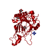
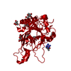
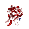
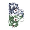
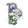
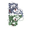
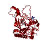
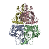
| Title | Crystal structure of Acinetobacter sp. DL28 L-ribose isomerase mutant E204Q in complex with L-ribulose | |||||||||
 Components Components | L-Ribose isomerase | |||||||||
 Keywords Keywords | ISOMERASE / Cupin barrel / Sugar binding | |||||||||
| Function / homology |  Function and homology information Function and homology informationRmlC-like cupin domain superfamily / Jelly Rolls / RmlC-like jelly roll fold / Jelly Rolls / Sandwich / Mainly Beta Similarity search - Domain/homology | |||||||||
| Biological species |  Acinetobacter (bacteria) Acinetobacter (bacteria) | |||||||||
| Method |  X-RAY DIFFRACTION / MOLECULAR REPLACEMENT / Resolution: 1.98 Å X-RAY DIFFRACTION / MOLECULAR REPLACEMENT / Resolution: 1.98 Å | |||||||||
 Authors Authors | Yoshida, H. / Yoshihara, A. / Teraoka, M. / Izumori, K. / Kamitori, S. | |||||||||
 Citation Citation | Journal: Febs J. / Year: 2014 Title: X-ray structure of a novel L-ribose isomerase acting on a non-natural sugar L-ribose as its ideal substrate. Authors: Yoshida, H. / Yoshihara, A. / Teraoka, M. / Terami, Y. / Takata, G. / Izumori, K. / Kamitori, S. #1: Journal: Acta Crystallogr.,Sect.F / Year: 2011 Title: Overexpression, crystallization and preliminary X-ray diffraction analysis of L-ribose isomerase from Acinetobacter sp. strain DL-28 Authors: Yoshida, H. / Teraoka, M. / Yoshihara, A. / Izumori, K. / Kamitori, S. | |||||||||
| History |
|
- Structure visualization
| Structure viewer | Molecule: MolmilJmol/JSmol |
|---|
- Downloads & links
-Download
| PDBx/mmCIF format | 4q0v.cif.gz | 67.4 KB | Display | PDBx/mmCIF format |
|---|---|---|---|---|
| PDB format | pdb4q0v.ent.gz | 48.3 KB | Display | PDB format |
| PDBx/mmJSON format | 4q0v.json.gz | Tree view | PDBx/mmJSON format | |
| Others |  Other downloads Other downloads |
-Validation report
| Arichive directory | https://data.pdbj.org/pub/pdb/validation_reports/q0/4q0vftp://data.pdbj.org/pub/pdb/validation_reports/q0/4q0v | HTTPS FTP |
|---|
-Related structure data
| Related structure data |  4q0pSC  4q0qC  4q0sC  4q0uC S: Starting model for refinement C: citing same article ( |
|---|---|
| Similar structure data |
-Links
 PDBj
PDBj
- Assembly
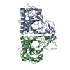
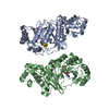
| Deposited unit | 
| ||||||||
|---|---|---|---|---|---|---|---|---|---|
| 1 | 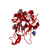
| ||||||||
| Unit cell |
|
-Components
-Protein , 1 types, 1 molecules A
| #1: Protein | Mass: 28750.270 Da / Num. of mol.: 1 / Mutation: E204Q Source method: isolated from a genetically manipulated source Source: (gene. exp.) Acinetobacter (bacteria) / Strain: DL-28 / Gene: L-RI / Plasmid: pQE30 / Production host:  Escherichia coli (E. coli) / Strain (production host): JM109 Escherichia coli (E. coli) / Strain (production host): JM109References: UniProt: Q93UQ5, Isomerases; Intramolecular oxidoreductases; Interconverting aldoses and ketoses, and related compounds |
|---|
-Sugars , 2 types, 7 molecules 

| #3: Sugar | 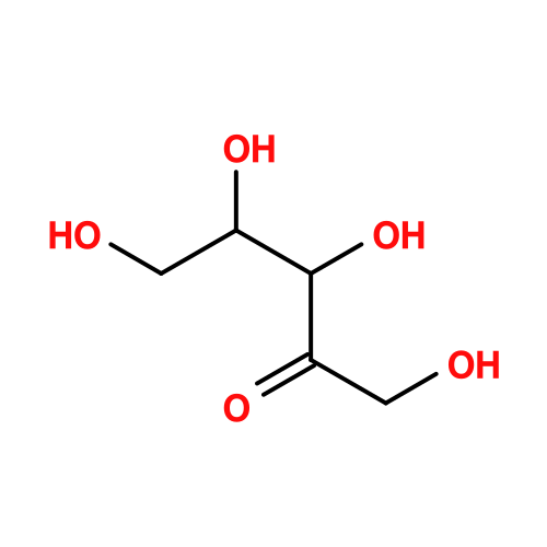 Type: L-saccharide / Mass: 150.130 Da / Num. of mol.: 2 Type: L-saccharide / Mass: 150.130 Da / Num. of mol.: 2Source method: isolated from a genetically manipulated source Formula: C5H10O5 #4: Sugar | ChemComp-RUU / 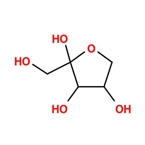 Type: L-saccharide, alpha linking / Mass: 150.130 Da / Num. of mol.: 5 Type: L-saccharide, alpha linking / Mass: 150.130 Da / Num. of mol.: 5Source method: isolated from a genetically manipulated source Formula: C5H10O5 |
|---|
-Non-polymers , 3 types, 103 molecules 

| #2: Chemical | ChemComp-CO /  Mass: 58.933 Da / Num. of mol.: 1 / Source method: obtained synthetically / Formula: Co Mass: 58.933 Da / Num. of mol.: 1 / Source method: obtained synthetically / Formula: Co |
|---|---|
| #5: Chemical | ChemComp-NCO /  Mass: 161.116 Da / Num. of mol.: 1 / Source method: obtained synthetically / Formula: CoH18N6 Mass: 161.116 Da / Num. of mol.: 1 / Source method: obtained synthetically / Formula: CoH18N6 |
| #6: Water | ChemComp-HOH / Mass: 18.015 Da / Num. of mol.: 101 / Source method: isolated from a natural source / Formula: H2O |
-Details
| Has protein modification | N |
|---|
-Experimental details
-Experiment
| Experiment | Method: X-RAY DIFFRACTION / Number of used crystals: 1 |
|---|
- Sample preparation
| Crystal | Density Matthews: 2.64 Å3/Da / Density % sol: 53.47 % |
|---|---|
| Crystal grow | Temperature: 293 K / Method: vapor diffusion, hanging drop / pH: 7.5 Details: 10-16%(v/v) PEG400, 0.2M NaCl, 0.1M HEPES, 0.02M Hexaammine cobalt(III) chloride, pH 7.5, VAPOR DIFFUSION, HANGING DROP, temperature 293K |
-Data collection
| Diffraction | Mean temperature: 100 K |
|---|---|
| Diffraction source | Source: ROTATING ANODE / Type: RIGAKU MICROMAX-007 HF / Wavelength: 1.5418 Å |
| Detector | Type: RIGAKU RAXIS VII / Detector: IMAGE PLATE / Date: Apr 12, 2012 / Details: Mirrors |
| Radiation | Protocol: SINGLE WAVELENGTH / Monochromatic (M) / Laue (L): M / Scattering type: x-ray |
| Radiation wavelength | Wavelength: 1.5418 Å / Relative weight: 1 |
| Reflection | Resolution: 1.98→21.8 Å / Num. all: 21360 / Num. obs: 21360 / % possible obs: 100 % / Observed criterion σ(F): 0 / Observed criterion σ(I): 0 / Redundancy: 6.5 % / Biso Wilson estimate: 20.9 Å2 / Rmerge(I) obs: 0.09 / Net I/σ(I): 10.6 |
| Reflection shell | Resolution: 1.98→2.05 Å / Redundancy: 6.4 % / Rmerge(I) obs: 0.404 / Mean I/σ(I) obs: 3.8 / Num. unique all: 2121 / % possible all: 100 |
- Processing
| Software |
| ||||||||||||||||||||||||||||||||||||
|---|---|---|---|---|---|---|---|---|---|---|---|---|---|---|---|---|---|---|---|---|---|---|---|---|---|---|---|---|---|---|---|---|---|---|---|---|---|
| Refinement | Method to determine structure: MOLECULAR REPLACEMENT Starting model: PDB ENTRY 4Q0P Resolution: 1.98→21.77 Å / Rfactor Rfree error: 0.005 / Data cutoff high absF: 2781687.42 / Data cutoff low absF: 0 / Isotropic thermal model: RESTRAINED / Cross valid method: THROUGHOUT / σ(F): 0 / σ(I): 0 / Stereochemistry target values: Engh & Huber / Details: BULK SOLVENT MODEL USED
| ||||||||||||||||||||||||||||||||||||
| Solvent computation | Solvent model: FLAT MODEL / Bsol: 70.3035 Å2 / ksol: 0.5 e/Å3 | ||||||||||||||||||||||||||||||||||||
| Displacement parameters | Biso mean: 33 Å2
| ||||||||||||||||||||||||||||||||||||
| Refine analyze |
| ||||||||||||||||||||||||||||||||||||
| Refinement step | Cycle: LAST / Resolution: 1.98→21.77 Å
| ||||||||||||||||||||||||||||||||||||
| Refine LS restraints |
| ||||||||||||||||||||||||||||||||||||
| LS refinement shell | Resolution: 1.98→2.05 Å / Rfactor Rfree error: 0.023 / Total num. of bins used: 10
| ||||||||||||||||||||||||||||||||||||
| Xplor file |
|
