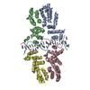
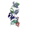
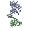
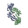
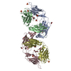
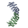
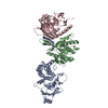
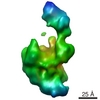
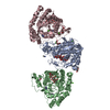
AUTHOR DETERMINED BIOLOGICAL UNIT IS TETRAMERIC. ONE IS DIMER PROTEIN (A,B) AND DOUBLE STRANDED DNA (E,F). THE OTHER IS DIMER PROTEIN (C,D) AND DOUBLE STRANDED DNA (E,F).
-
Components
#1: Protein
CprB
Mass: 23719.328 Da / Num. of mol.: 4 Source method: isolated from a genetically manipulated source Source: (gene. exp.) Streptomyces coelicolor (bacteria) / Gene: cprB / Production host: Escherichia coli (E. coli) / References: UniProt: O66122
#2: DNA chain
DNA (5'-D(*AP*CP*AP*TP*AP*CP*GP*GP*GP*AP*CP*GP*CP*CP*CP*CP*GP*TP*TP*TP*AP*T)-3')
Resolution: 3.2→60.9 Å / Cor.coef. Fo:Fc: 0.933 / Cor.coef. Fo:Fc free: 0.884 / SU B: 57.729 / SU ML: 0.425 / Cross valid method: THROUGHOUT / ESU R Free: 0.483 / Stereochemistry target values: MAXIMUM LIKELIHOOD / Details: HYDROGENS HAVE BEEN USED IF PRESENT IN THE INPUT
Rfactor
Num. reflection
% reflection
Selection details
Rfree
0.29433
1163
4.1 %
RANDOM
Rwork
0.21985
-
-
-
obs
0.22299
27073
99.56 %
-
all
-
28269
-
-
Solvent computation
Ion probe radii: 0.8 Å / Shrinkage radii: 0.8 Å / VDW probe radii: 1.2 Å / Solvent model: MASK
 Movie
Movie Controller

 Yorodumi
 Open data
 Basic information
 Components
 Keywords
 Function and homology information
 Streptomyces coelicolor (bacteria)
 X-RAY DIFFRACTION /
  Authors
 Citation
 Structure visualization
 Downloads & links
 Other downloads
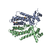

 PDBj
PDBj


 Assembly
 Sample preparation
 / Beamline: BM14 / Wavelength: 0.98 Å
 Processing