- PDB-4pxg: Crystal Structure of TypeII restriction Enzyme Sau3AI -
+
Open data
ID or keywords:
Loading...
-
Basic information
Entry
Database: PDB / ID: 4pxg
Title
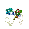
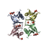
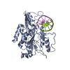
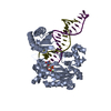
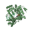
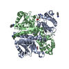
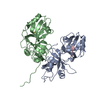
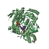
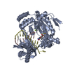
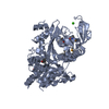
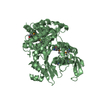
Crystal Structure of TypeII restriction Enzyme Sau3AI
Components
Type-2 restriction enzyme Sau3AI
Keywords
HYDROLASE / Type II restriction enzyme
Function / homology
Function and homology information
type II site-specific deoxyribonuclease / type II site-specific deoxyribonuclease activity / DNA restriction-modification system / DNA binding Similarity search - Function
DNA mismatch repair MutH/Type II restriction enzyme Sau3AI / DNA mismatch repair enzyme MutH / DNA mismatch repair enzyme MutH / DNA mismatch repair MutH/Type II restriction endonuclease superfamily / Restriction endonuclease type II-like Similarity search - Domain/homology
Method to determine structure: SAD / Resolution: 2.45→50 Å / Cor.coef. Fo:Fc: 0.939 / Cor.coef. Fo:Fc free: 0.914 / SU B: 11.307 / SU ML: 0.245 / Cross valid method: THROUGHOUT / ESU R: 0.432 / ESU R Free: 0.28 / Stereochemistry target values: MAXIMUM LIKELIHOOD / Details: HYDROGENS HAVE BEEN ADDED IN THE RIDING POSITIONS
Rfactor
Num. reflection
% reflection
Selection details
Rfree
0.26331
2331
5.1 %
RANDOM
Rwork
0.22373
-
-
-
obs
0.22576
43797
94.07 %
-
all
-
46127
-
-
Solvent computation
Ion probe radii: 0.8 Å / Shrinkage radii: 0.8 Å / VDW probe radii: 1.2 Å / Solvent model: MASK
 Movie
Movie Controller


 Open data
 Basic information
 Components
 Keywords
 Function and homology information
 Staphylococcus aureus (bacteria)
 X-RAY DIFFRACTION /
  Authors
 Citation
 Structure visualization
 Downloads & links
 Other downloads
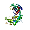

 PDBj
PDBj


 Assembly

 Mass: 40.078 Da / Num. of mol.: 1 / Source method: obtained synthetically / Formula: Ca
 Mass: 18.015 Da / Num. of mol.: 31 / Source method: isolated from a natural source / Formula: H2O
 Sample preparation
 Processing