 Movie
Movie Controller


+ Open data

- Basic information
| Entry | Database: PDB / ID: 4psn | ||||||
|---|---|---|---|---|---|---|---|
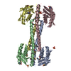
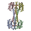
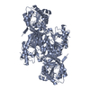
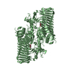
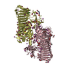
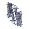
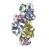
| Title | Crystal structure of apeThermo-DBP-RP2 | ||||||
 Components Components | ssDNA binding protein | ||||||
 Keywords Keywords | DNA BINDING PROTEIN / ssDNA binding protein | ||||||
| Function / homology | : / Thermo-DBP-RP2 C-terminal domain / ThermoDBP-related, archaea / ThermoDBP-related, archaea / IMIDAZOLE / Thermo-DBP-RP2-like C-terminal domain-containing protein Function and homology information Function and homology information | ||||||
| Biological species |   Aeropyrum pernix (archaea) Aeropyrum pernix (archaea) | ||||||
| Method |  X-RAY DIFFRACTION / SYNCHROTRON / SAD / Resolution: 2.05 Å X-RAY DIFFRACTION / SYNCHROTRON / SAD / Resolution: 2.05 Å | ||||||
 Authors Authors | Gahlei, H. / von Moeller, H. / Eppers, D. / Loll, B. / Wahl, M.C. | ||||||
 Citation Citation | Journal: Nucleic Acids Res. / Year: 2014 Title: Entrapment of DNA in an intersubunit tunnel system of a single-stranded DNA-binding protein. Authors: Ghalei, H. / Moeller, H.v. / Eppers, D. / Sohmen, D. / Wilson, D.N. / Loll, B. / Wahl, M.C. | ||||||
| History |
|
- Structure visualization
| Structure viewer | Molecule: MolmilJmol/JSmol |
|---|
- Downloads & links
-Download
| PDBx/mmCIF format | 4psn.cif.gz | 365.3 KB | Display | PDBx/mmCIF format |
|---|---|---|---|---|
| PDB format | pdb4psn.ent.gz | 301.2 KB | Display | PDB format |
| PDBx/mmJSON format | 4psn.json.gz | Tree view | PDBx/mmJSON format | |
| Others |  Other downloads Other downloads |
-Validation report
| Arichive directory | https://data.pdbj.org/pub/pdb/validation_reports/ps/4psnftp://data.pdbj.org/pub/pdb/validation_reports/ps/4psn | HTTPS FTP |
|---|
-Related structure data


-Links
 PDBj
PDBj


- Assembly
| Deposited unit | 
| ||||||||
|---|---|---|---|---|---|---|---|---|---|
| 1 |
| ||||||||
| Unit cell |
|
-Components
| #1: Protein | Mass: 26768.100 Da / Num. of mol.: 4 Source method: isolated from a genetically manipulated source Source: (gene. exp.) Aeropyrum pernix (archaea)Strain: ATCC 700893 / DSM 11879 / JCM 9820 / NBRC 100138 / K1 Gene: APE_1866.1 / Plasmid: pET-M11 / Production host:  #2: Chemical | ChemComp-IMD /   Mass: 69.085 Da / Num. of mol.: 6 / Source method: obtained synthetically / Formula: C3H5N2 Mass: 69.085 Da / Num. of mol.: 6 / Source method: obtained synthetically / Formula: C3H5N2#3: Chemical |   Mass: 92.094 Da / Num. of mol.: 2 / Source method: obtained synthetically / Formula: C3H8O3 Mass: 92.094 Da / Num. of mol.: 2 / Source method: obtained synthetically / Formula: C3H8O3#4: Water | ChemComp-HOH / |  Mass: 18.015 Da / Num. of mol.: 283 / Source method: isolated from a natural source / Formula: H2O Mass: 18.015 Da / Num. of mol.: 283 / Source method: isolated from a natural source / Formula: H2OHas protein modification | Y | |
|---|
-Experimental details
-Experiment
| Experiment | Method: X-RAY DIFFRACTION / Number of used crystals: 1 |
|---|
- Sample preparation
| Crystal | Density Matthews: 2.41 Å3/Da / Density % sol: 48.88 % |
|---|---|
| Crystal grow | Temperature: 293 K / Method: vapor diffusion, sitting drop / pH: 8 Details: 0.1 M imidazole, pH 8.0, 0.4 M NaH2PO4, 1.6 M K2HPO4, 0.2 M NaCl and 0.25 M glycine, VAPOR DIFFUSION, SITTING DROP, temperature 293K |
-Data collection
| Diffraction | Mean temperature: 100 K |
|---|---|
| Diffraction source | Source: SYNCHROTRON / Site: BESSY  / Beamline: 14.2 / Wavelength: 0.98 Å / Beamline: 14.2 / Wavelength: 0.98 Å |
| Detector | Type: RAYONIX MX-225 / Detector: CCD / Date: May 7, 2010 |
| Radiation | Monochromator: SAGITALLY FOCUSED SI(111) / Protocol: SINGLE WAVELENGTH / Monochromatic (M) / Laue (L): M / Scattering type: x-ray |
| Radiation wavelength | Wavelength: 0.98 Å / Relative weight: 1 |
| Reflection | Resolution: 2.05→50 Å / Num. all: 125145 / % possible obs: 99.9 % / Observed criterion σ(I): -3 / Redundancy: 7.4 % / Biso Wilson estimate: 28.8 Å2 |
| Reflection shell | Resolution: 2.05→2.1 Å / Redundancy: 5.9 % / % possible all: 100 |
- Processing
| Software |
| ||||||||||||||||||||||||||||||||||||||||||||||||||||||||||||||||||||||||||||||||||||||||||||||||||||||||||||||||||||||||||||||||||||||||||||||||||||||||||||||||||||||||
|---|---|---|---|---|---|---|---|---|---|---|---|---|---|---|---|---|---|---|---|---|---|---|---|---|---|---|---|---|---|---|---|---|---|---|---|---|---|---|---|---|---|---|---|---|---|---|---|---|---|---|---|---|---|---|---|---|---|---|---|---|---|---|---|---|---|---|---|---|---|---|---|---|---|---|---|---|---|---|---|---|---|---|---|---|---|---|---|---|---|---|---|---|---|---|---|---|---|---|---|---|---|---|---|---|---|---|---|---|---|---|---|---|---|---|---|---|---|---|---|---|---|---|---|---|---|---|---|---|---|---|---|---|---|---|---|---|---|---|---|---|---|---|---|---|---|---|---|---|---|---|---|---|---|---|---|---|---|---|---|---|---|---|---|---|---|---|---|---|---|
| Refinement | Method to determine structure: SAD / Resolution: 2.05→32.727 Å / SU ML: 0.25 / Phase error: 20.27 / Stereochemistry target values: ML
| ||||||||||||||||||||||||||||||||||||||||||||||||||||||||||||||||||||||||||||||||||||||||||||||||||||||||||||||||||||||||||||||||||||||||||||||||||||||||||||||||||||||||
| Solvent computation | Shrinkage radii: 0.83 Å / VDW probe radii: 1.1 Å / Solvent model: FLAT BULK SOLVENT MODEL / Bsol: 53.614 Å2 / ksol: 0.419 e/Å3 | ||||||||||||||||||||||||||||||||||||||||||||||||||||||||||||||||||||||||||||||||||||||||||||||||||||||||||||||||||||||||||||||||||||||||||||||||||||||||||||||||||||||||
| Displacement parameters |
| ||||||||||||||||||||||||||||||||||||||||||||||||||||||||||||||||||||||||||||||||||||||||||||||||||||||||||||||||||||||||||||||||||||||||||||||||||||||||||||||||||||||||
| Refinement step | Cycle: LAST / Resolution: 2.05→32.727 Å
| ||||||||||||||||||||||||||||||||||||||||||||||||||||||||||||||||||||||||||||||||||||||||||||||||||||||||||||||||||||||||||||||||||||||||||||||||||||||||||||||||||||||||
| Refine LS restraints |
| ||||||||||||||||||||||||||||||||||||||||||||||||||||||||||||||||||||||||||||||||||||||||||||||||||||||||||||||||||||||||||||||||||||||||||||||||||||||||||||||||||||||||
| LS refinement shell |
| ||||||||||||||||||||||||||||||||||||||||||||||||||||||||||||||||||||||||||||||||||||||||||||||||||||||||||||||||||||||||||||||||||||||||||||||||||||||||||||||||||||||||
| Refinement TLS params. | Method: refined / Origin x: -15.3485 Å / Origin y: -15.9074 Å / Origin z: 38.1118 Å
| ||||||||||||||||||||||||||||||||||||||||||||||||||||||||||||||||||||||||||||||||||||||||||||||||||||||||||||||||||||||||||||||||||||||||||||||||||||||||||||||||||||||||
| Refinement TLS group | Selection details: all |
