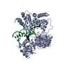
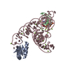
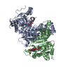
- PDB-4pht: ATPase GspE in complex with the cytoplasmic domain of GspL from t... -
+
Open data
ID or keywords:
Loading...
-
Basic information
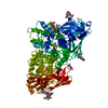
Entry
Database: PDB / ID: 4pht
Title
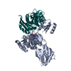
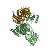
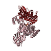
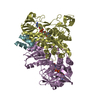
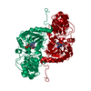
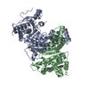
ATPase GspE in complex with the cytoplasmic domain of GspL from the Vibrio vulnificus type II Secretion system
Components
General secretory pathway protein E
Type II secretion system protein L
Keywords
PROTEIN TRANSPORT
Function / homology
Function and homology information
Gram-negative-bacterium-type cell wall / protein secretion by the type II secretion system / type II protein secretion system complex / membrane => GO:0016020 / nucleotide binding / ATP hydrolysis activity / ATP binding / metal ion binding / plasma membrane / cytoplasm Similarity search - Function
: / Type II secretion system, protein E, N-terminal domain / GspL cytoplasmic domain, C-terminal subdomain / Nucleotidyltransferase; domain 5 - #380 / : / GSPII protein E, N1E domain / Type II secretion system protein GspE / Type II secretion system protein GspL / GspL, cytoplasmic actin-ATPase-like domain / GspL periplasmic domain ...: / Type II secretion system, protein E, N-terminal domain / GspL cytoplasmic domain, C-terminal subdomain / Nucleotidyltransferase; domain 5 - #380 / : / GSPII protein E, N1E domain / Type II secretion system protein GspE / Type II secretion system protein GspL / GspL, cytoplasmic actin-ATPase-like domain / GspL periplasmic domain / Type II secretion system (T2SS), protein L / GspL periplasmic domain / Beta-Lactamase - #90 / Type II secretion system protein GspE, N-terminal superfamily / Bacterial type II secretion system protein E signature. / Type II/IV secretion system protein / Type II/IV secretion system protein / GMP Synthetase; Chain A, domain 3 / Beta-Lactamase / ATPase, nucleotide binding domain / Nucleotidyltransferase; domain 5 / P-loop containing nucleotide triphosphate hydrolases / ATPases associated with a variety of cellular activities / AAA+ ATPase domain / Rossmann fold / P-loop containing nucleoside triphosphate hydrolase / 2-Layer Sandwich / 3-Layer(aba) Sandwich / Alpha Beta Similarity search - Domain/homology
PHOSPHOAMINOPHOSPHONIC ACID-ADENYLATE ESTER / Type II secretion system protein L / Type II secretion system protein E / Type II secretion system protein E / Type II secretion system protein L Similarity search - Component
Biological species
Vibrio vulnificus (bacteria)
Method
X-RAY DIFFRACTION / SYNCHROTRON / Resolution: 2.83 Å
#95 - Nov 2007 Multidrug Resistance Transporters similarity (1)
-
Assembly
Deposited unit
X: Type II secretion system protein L A: General secretory pathway protein E Y: Type II secretion system protein L B: General secretory pathway protein E Z: Type II secretion system protein L C: General secretory pathway protein E hetero molecules
 Movie
Movie Controller

 Yorodumi
 Open data
 Basic information
 Components
 Keywords
 Function and homology information
 Vibrio vulnificus (bacteria)
 X-RAY DIFFRACTION /
  Authors
 United States, 1items
  Citation
 Structure visualization
 Downloads & links
 Other downloads
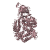
 PDBj
PDBj


 Assembly