 Movie
Movie Controller

[English] 日本語
 Yorodumi
- PDB-2dht: Crystal structure of isocitrate dehydrogenase from Sulfolobus tok... -
+ Open data

- Basic information
| Entry | Database: PDB / ID: 2dht | ||||||
|---|---|---|---|---|---|---|---|
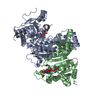
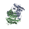
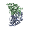
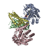
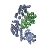
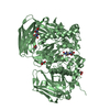
| Title | Crystal structure of isocitrate dehydrogenase from Sulfolobus tokodaii strain7 | ||||||
 Components Components | 409aa long hypothetical NADP-dependent isocitrate dehydrogenase | ||||||
 Keywords Keywords | OXIDOREDUCTASE / homo dimer | ||||||
| Function / homology |  Function and homology information Function and homology informationisocitrate dehydrogenase (NADP+) / isocitrate dehydrogenase (NADP+) activity / tricarboxylic acid cycle / nucleotide binding / metal ion binding Similarity search - Function | ||||||
| Biological species |   Sulfolobus tokodaii (archaea) Sulfolobus tokodaii (archaea) | ||||||
| Method |  X-RAY DIFFRACTION / SYNCHROTRON / MOLECULAR REPLACEMENT / Resolution: 2.5 Å X-RAY DIFFRACTION / SYNCHROTRON / MOLECULAR REPLACEMENT / Resolution: 2.5 Å | ||||||
 Authors Authors | Kondo, H. / Murakami, M. / Ihara, K. / Suzuki, S. / Kouyama, T. | ||||||
 Citation Citation | Journal: To be Published Title: Crystal structure of isocitrate dehydrogenase of Sulfolobus tokodaii strain7 Authors: Kondo, H. / Murakami, M. / Ihara, K. / Suzuki, S. / Kouyama, T. | ||||||
| History |
|
- Structure visualization
| Structure viewer | Molecule: MolmilJmol/JSmol |
|---|
- Downloads & links
-Download
| PDBx/mmCIF format | 2dht.cif.gz | 167.7 KB | Display | PDBx/mmCIF format |
|---|---|---|---|---|
| PDB format | pdb2dht.ent.gz | 133.4 KB | Display | PDB format |
| PDBx/mmJSON format | 2dht.json.gz | Tree view | PDBx/mmJSON format | |
| Others |  Other downloads Other downloads |
-Validation report
| Arichive directory | https://data.pdbj.org/pub/pdb/validation_reports/dh/2dhtftp://data.pdbj.org/pub/pdb/validation_reports/dh/2dht | HTTPS FTP |
|---|
-Related structure data
| Related structure data |  1v94S S: Starting model for refinement |
|---|---|
| Similar structure data |


-Links
 PDBj
PDBj

- Assembly
| Deposited unit | 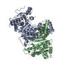
| ||||||||
|---|---|---|---|---|---|---|---|---|---|
| 1 |
| ||||||||
| Unit cell |
|
-Components
| #1: Protein | Mass: 46558.871 Da / Num. of mol.: 2 Source method: isolated from a genetically manipulated source Source: (gene. exp.) Sulfolobus tokodaii (archaea) / Strain: str. 7 / Production host:  References: GenBank: 15623283, UniProt: Q96YK6*PLUS, isocitrate dehydrogenase (NADP+) #2: Water | ChemComp-HOH / |  Mass: 18.015 Da / Num. of mol.: 38 / Source method: isolated from a natural source / Formula: H2O Mass: 18.015 Da / Num. of mol.: 38 / Source method: isolated from a natural source / Formula: H2O |
|---|
-Experimental details
-Experiment
| Experiment | Method: X-RAY DIFFRACTION / Number of used crystals: 1 |
|---|
- Sample preparation
| Crystal | Density Matthews: 2.66 Å3/Da / Density % sol: 53.82 % |
|---|---|
| Crystal grow | Temperature: 283 K / Method: vapor diffusion, hanging drop / pH: 7.5 Details: 14% PEG 10000, 0.1M HEPES, pH 7.5, VAPOR DIFFUSION, HANGING DROP, temperature 283K |
-Data collection
| Diffraction | Mean temperature: 100 K |
|---|---|
| Diffraction source | Source: SYNCHROTRON / Site: SPring-8  / Beamline: BL41XU / Wavelength: 1 Å / Beamline: BL41XU / Wavelength: 1 Å |
| Detector | Type: RIGAKU / Detector: IMAGE PLATE / Date: Sep 30, 2005 |
| Radiation | Monochromator: double-crystal monochomator / Protocol: SINGLE WAVELENGTH / Monochromatic (M) / Laue (L): M / Scattering type: x-ray |
| Radiation wavelength | Wavelength: 1 Å / Relative weight: 1 |
| Reflection | Resolution: 2.5→53.6 Å / Num. all: 33797 / Num. obs: 33300 / % possible obs: 97.5 % / Observed criterion σ(F): 1.4 / Observed criterion σ(I): 1.4 / Redundancy: 3.2 % / Biso Wilson estimate: 61.5 Å2 / Rmerge(I) obs: 0.061 / Rsym value: 0.061 / Net I/σ(I): 10.6 |
| Reflection shell | Resolution: 2.5→2.64 Å / Redundancy: 3.4 % / Rmerge(I) obs: 0.771 / Mean I/σ(I) obs: 1.6 / Num. unique all: 4511 / Rsym value: 0.492 / % possible all: 98.7 |
- Processing
| Software |
| ||||||||||||||||||||||||||||||||||||||||||||||||||||||||||||||||||||||||||||||||||||||||||
|---|---|---|---|---|---|---|---|---|---|---|---|---|---|---|---|---|---|---|---|---|---|---|---|---|---|---|---|---|---|---|---|---|---|---|---|---|---|---|---|---|---|---|---|---|---|---|---|---|---|---|---|---|---|---|---|---|---|---|---|---|---|---|---|---|---|---|---|---|---|---|---|---|---|---|---|---|---|---|---|---|---|---|---|---|---|---|---|---|---|---|---|
| Refinement | Method to determine structure: MOLECULAR REPLACEMENT Starting model: 1v94 Resolution: 2.5→15 Å / Rfactor Rfree error: 0.007 / Occupancy max: 1 / Occupancy min: 1 / Isotropic thermal model: anisotropic / Cross valid method: THROUGHOUT / σ(F): 0 / Stereochemistry target values: Engh & Huber
| ||||||||||||||||||||||||||||||||||||||||||||||||||||||||||||||||||||||||||||||||||||||||||
| Solvent computation | Solvent model: CNS bulk solvent model used / Bsol: 39.5232 Å2 / ksol: 0.333597 e/Å3 | ||||||||||||||||||||||||||||||||||||||||||||||||||||||||||||||||||||||||||||||||||||||||||
| Displacement parameters | Biso max: 121.83 Å2 / Biso mean: 55.96 Å2 / Biso min: 20.26 Å2
| ||||||||||||||||||||||||||||||||||||||||||||||||||||||||||||||||||||||||||||||||||||||||||
| Refine analyze |
| ||||||||||||||||||||||||||||||||||||||||||||||||||||||||||||||||||||||||||||||||||||||||||
| Refinement step | Cycle: LAST / Resolution: 2.5→15 Å
| ||||||||||||||||||||||||||||||||||||||||||||||||||||||||||||||||||||||||||||||||||||||||||
| Refine LS restraints |
| ||||||||||||||||||||||||||||||||||||||||||||||||||||||||||||||||||||||||||||||||||||||||||
| LS refinement shell | Refine-ID: X-RAY DIFFRACTION / Total num. of bins used: 8
| ||||||||||||||||||||||||||||||||||||||||||||||||||||||||||||||||||||||||||||||||||||||||||
| Xplor file |
|
