 Movie
Movie Controller

[English] 日本語
 Yorodumi
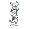
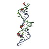
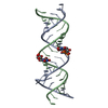
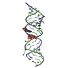
- PDB-4pdq: Crystal structure of the bacterial ribosomal decoding site in com... -
+ Open data

- Basic information
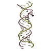
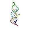
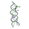
| Entry | Database: PDB / ID: 4pdq | ||||||||||||||||||||
|---|---|---|---|---|---|---|---|---|---|---|---|---|---|---|---|---|---|---|---|---|---|
| Title | Crystal structure of the bacterial ribosomal decoding site in complex with 4'-deoxy-4'-fluoro neomycin analog | ||||||||||||||||||||
 Components Components | RNA (5'-* Keywords KeywordsRNA/ANTIBIOTIC / ribosome / aminoglycoside / RNA-ANTIBIOTIC complex | Function / homology | Chem-NMZ / RNA / RNA (> 10) |  Function and homology information Function and homology informationBiological species | synthetic (others) | Method |  X-RAY DIFFRACTION / SYNCHROTRON / MOLECULAR REPLACEMENT / molecular replacement / Resolution: 3 Å X-RAY DIFFRACTION / SYNCHROTRON / MOLECULAR REPLACEMENT / molecular replacement / Resolution: 3 Å  Authors AuthorsHanessian, S. / Saavedra, O.M. / Vilchis-Reyes, M.A. / Maianti, J.P. / Kanazawa, H. / Dozzo, P. / Feeney, L.A. / Armstrong, E.S. / Kondo, J. |  CitationJournal: Chem Sci / Year: 2014 CitationJournal: Chem Sci / Year: 2014Title: Synthesis, broad spectrum antibacterial activity, and X-ray co-crystal structure of the decoding bacterial ribosomal A-site with 4'-deoxy-4'-fluoro neomycin analogs Authors: Hanessian, S. / Saavedra, O.M. / Vilchis-Reyes, M.A. / Maianti, J.P. / Kanazawa, H. / Dozzo, P. / Matias, R.D. / Serio, A. / Kondo, J. History |
|
- Structure visualization
| Structure viewer | Molecule: MolmilJmol/JSmol |
|---|
- Downloads & links
-Download
| PDBx/mmCIF format | 4pdq.cif.gz | 36.4 KB | Display | PDBx/mmCIF format |
|---|---|---|---|---|
| PDB format | pdb4pdq.ent.gz | 25 KB | Display | PDB format |
| PDBx/mmJSON format | 4pdq.json.gz | Tree view | PDBx/mmJSON format | |
| Others |  Other downloads Other downloads |
-Validation report
| Arichive directory | https://data.pdbj.org/pub/pdb/validation_reports/pd/4pdqftp://data.pdbj.org/pub/pdb/validation_reports/pd/4pdq | HTTPS FTP |
|---|
-Related structure data
| Similar structure data |
|---|


-Links
 PDBj
PDBj


- Assembly
| Deposited unit | 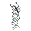
| ||||||||
|---|---|---|---|---|---|---|---|---|---|
| 1 |
| ||||||||
| Unit cell |
|
-Components
| #1: RNA chain | Mass: 7370.424 Da / Num. of mol.: 2 / Source method: obtained synthetically / Source: (synth.) synthetic (others) #2: Chemical |   Mass: 24.305 Da / Num. of mol.: 2 / Source method: obtained synthetically / Formula: Mg Mass: 24.305 Da / Num. of mol.: 2 / Source method: obtained synthetically / Formula: Mg#3: Chemical | ChemComp-NMZ / ( | 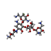  Mass: 717.739 Da / Num. of mol.: 1 / Source method: obtained synthetically / Formula: C27H52FN7O14 Mass: 717.739 Da / Num. of mol.: 1 / Source method: obtained synthetically / Formula: C27H52FN7O14#4: Water | ChemComp-HOH / |  Mass: 18.015 Da / Num. of mol.: 1 / Source method: isolated from a natural source / Formula: H2O Mass: 18.015 Da / Num. of mol.: 1 / Source method: isolated from a natural source / Formula: H2O |
|---|
-Experimental details
-Experiment
| Experiment | Method: X-RAY DIFFRACTION / Number of used crystals: 1 |
|---|
- Sample preparation
| Crystal | Density Matthews: 3.13 Å3/Da / Density % sol: 60.66 % |
|---|---|
| Crystal grow | Temperature: 293 K / Method: vapor diffusion, sitting drop / pH: 7 Details: Sodium cacodylate, MgCl2, MPD, Spermine tetrahydrochloride |
-Data collection
| Diffraction | Mean temperature: 100 K |
|---|---|
| Diffraction source | Source: SYNCHROTRON / Site: Photon Factory  / Beamline: BL-17A / Wavelength: 0.98 Å / Beamline: BL-17A / Wavelength: 0.98 Å |
| Detector | Type: ADSC QUANTUM 270 / Detector: CCD / Date: Nov 18, 2013 |
| Radiation | Protocol: SINGLE WAVELENGTH / Monochromatic (M) / Laue (L): M / Scattering type: x-ray |
| Radiation wavelength | Wavelength: 0.98 Å / Relative weight: 1 |
| Reflection | Resolution: 3→19.6 Å / Num. obs: 3522 / % possible obs: 98.7 % / Redundancy: 5.6 % / Net I/σ(I): 12.6 |
-Phasing
| Phasing | Method: molecular replacement |
|---|
- Processing
| Software |
| ||||||||||||||||||||||||||||||||||||||||||||||||||||||||||||||||||||||||||||||||
|---|---|---|---|---|---|---|---|---|---|---|---|---|---|---|---|---|---|---|---|---|---|---|---|---|---|---|---|---|---|---|---|---|---|---|---|---|---|---|---|---|---|---|---|---|---|---|---|---|---|---|---|---|---|---|---|---|---|---|---|---|---|---|---|---|---|---|---|---|---|---|---|---|---|---|---|---|---|---|---|---|---|
| Refinement | Method to determine structure: MOLECULAR REPLACEMENT / Resolution: 3→19.6 Å / Cross valid method: FREE R-VALUE / σ(F): 0
| ||||||||||||||||||||||||||||||||||||||||||||||||||||||||||||||||||||||||||||||||
| Solvent computation | Bsol: 59.88 Å2 | ||||||||||||||||||||||||||||||||||||||||||||||||||||||||||||||||||||||||||||||||
| Displacement parameters | Biso mean: 76.3504 Å2
| ||||||||||||||||||||||||||||||||||||||||||||||||||||||||||||||||||||||||||||||||
| Refinement step | Cycle: LAST / Resolution: 3→19.6 Å
| ||||||||||||||||||||||||||||||||||||||||||||||||||||||||||||||||||||||||||||||||
| Refine LS restraints |
| ||||||||||||||||||||||||||||||||||||||||||||||||||||||||||||||||||||||||||||||||
| LS refinement shell | Resolution: 3→3.11 Å
| ||||||||||||||||||||||||||||||||||||||||||||||||||||||||||||||||||||||||||||||||
| Xplor file |
|
