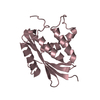
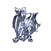
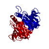
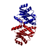
Entry Database : PDB / ID : 4pa1Title Crystal Structure of Catalytic Core domain of FIV Integrase Integrase Keywords / / / Function / homology Function Domain/homology Component
/ / / / / / / / / / / / / / / / / / / / / / / / / / / / / / / / / / / / / / / / / / / / / / / / / / / / / / / / / / / / / / / / / / / / / / / / / / / / / / / / / Biological species Method / / Resolution : 1.84 Å Authors Galilee, M. / Alian, A. Journal : Structure / Year : 2014Title : Identification of phe187 as a crucial dimerization determinant facilitates crystallization of a monomeric retroviral integrase core domain.Authors : Galilee, M. / Alian, A. History Deposition Apr 7, 2014 Deposition site / Processing site Revision 1.0 Sep 17, 2014 Provider / Type Revision 1.1 Oct 22, 2014 Group Revision 1.2 Dec 27, 2023 Group Advisory / Data collection ... Advisory / Data collection / Database references / Derived calculations / Other / Refinement description / Source and taxonomy / Structure summary Category chem_comp_atom / chem_comp_bond ... chem_comp_atom / chem_comp_bond / citation / database_2 / entity_src_gen / pdbx_database_related / pdbx_database_status / pdbx_struct_assembly / pdbx_struct_assembly_prop / pdbx_struct_oper_list / pdbx_validate_symm_contact / refine_hist / struct_keywords Item _citation.journal_id_CSD / _database_2.pdbx_DOI ... _citation.journal_id_CSD / _database_2.pdbx_DOI / _database_2.pdbx_database_accession / _entity_src_gen.pdbx_alt_source_flag / _pdbx_database_related.content_type / _pdbx_database_status.pdb_format_compatible / _pdbx_struct_assembly.oligomeric_details / _pdbx_struct_assembly_prop.type / _pdbx_struct_assembly_prop.value / _pdbx_struct_oper_list.symmetry_operation / _pdbx_validate_symm_contact.auth_seq_id_1 / _pdbx_validate_symm_contact.auth_seq_id_2 / _pdbx_validate_symm_contact.dist / _refine_hist.number_atoms_solvent / _refine_hist.number_atoms_total / _refine_hist.pdbx_number_atoms_ligand / _refine_hist.pdbx_number_atoms_nucleic_acid / _refine_hist.pdbx_number_atoms_protein / _struct_keywords.text Revision 1.3 Mar 27, 2024 Group / Category
Show all Show less
 Movie
Movie Controller


 Open data
 Basic information
 Components
 Keywords
 Function and homology information
 Feline immunodeficiency virus
 X-RAY DIFFRACTION /
  Authors
 Citation
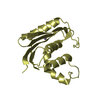
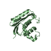
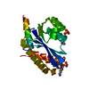
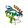
 Structure visualization
 Downloads & links
 Other downloads
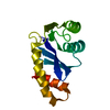


 PDBj
PDBj

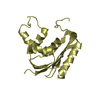
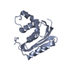
 Assembly

 Mass: 18.015 Da / Num. of mol.: 111 / Source method: isolated from a natural source / Formula: H2O
 Sample preparation
 Processing