 Movie
Movie Controller


+ Open data

- Basic information
| Entry | Database: PDB / ID: 4p9p | |||||||||
|---|---|---|---|---|---|---|---|---|---|---|
| Title | Structure of NavMS in complex with channel blocking compound | |||||||||
 Components Components | Ion transport protein | |||||||||
 Keywords Keywords | TRANSPORT PROTEIN / channel blocking compound / sodium channel / pore / membrane protein | |||||||||
| Function / homology |  Function and homology information Function and homology informationvoltage-gated sodium channel complex / voltage-gated sodium channel activity Similarity search - Function | |||||||||
| Biological species |  Magnetococcus sp. (bacteria) Magnetococcus sp. (bacteria) | |||||||||
| Method |  X-RAY DIFFRACTION / SYNCHROTRON / MOLECULAR REPLACEMENT / Resolution: 2.91 Å X-RAY DIFFRACTION / SYNCHROTRON / MOLECULAR REPLACEMENT / Resolution: 2.91 Å | |||||||||
 Authors Authors | Naylor, C.E. / Bagneris, C. / Wallace, B.A. | |||||||||
| Funding support |  United Kingdom, 2items United Kingdom, 2items
| |||||||||
 Citation Citation | Journal: Proc.Natl.Acad.Sci.USA / Year: 2014 Title: Prokaryotic NavMs channel as a structural and functional model for eukaryotic sodium channel antagonism. Authors: Bagneris, C. / DeCaen, P.G. / Naylor, C.E. / Pryde, D.C. / Nobeli, I. / Clapham, D.E. / Wallace, B.A. #1: Journal: Nat Commun / Year: 2013Title: Role of the C-terminal domain in the structure and function of tetrameric sodium channels. Authors: Bagneris, C. / Decaen, P.G. / Hall, B.A. / Naylor, C.E. / Clapham, D.E. / Kay, C.W. / Wallace, B.A. #2: Journal: Nat Commun / Year: 2012Title: Structure of a bacterial voltage-gated sodium channel pore reveals mechanisms of opening and closing. Authors: McCusker, E.C. / Bagneris, C. / Naylor, C.E. / Cole, A.R. / D'Avanzo, N. / Nichols, C.G. / Wallace, B.A. | |||||||||
| History |
| |||||||||
| Remark 0 | : statistics at the very beginning when nothing is done yet |
- Structure visualization
| Structure viewer | Molecule: MolmilJmol/JSmol |
|---|
- Downloads & links
-Download
| PDBx/mmCIF format | 4p9p.cif.gz | 170.7 KB | Display | PDBx/mmCIF format |
|---|---|---|---|---|
| PDB format | pdb4p9p.ent.gz | 133.8 KB | Display | PDB format |
| PDBx/mmJSON format | 4p9p.json.gz | Tree view | PDBx/mmJSON format | |
| Others |  Other downloads Other downloads |
-Validation report
| Arichive directory | https://data.pdbj.org/pub/pdb/validation_reports/p9/4p9pftp://data.pdbj.org/pub/pdb/validation_reports/p9/4p9p | HTTPS FTP |
|---|
-Related structure data
| Related structure data |  4cbcC  4oxsC  4p2zC  4p30C  4p9oC  4pa3C  4pa4C  4pa6C  4pa7C  4pa9C C: citing same article ( |
|---|---|
| Similar structure data |


-Links
 PDBj
PDBj


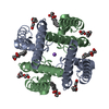
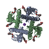
- Assembly
| Deposited unit | 
| ||||||||||||
|---|---|---|---|---|---|---|---|---|---|---|---|---|---|
| 1 | 
| ||||||||||||
| 2 | 
| ||||||||||||
| Unit cell |
| ||||||||||||
| Components on special symmetry positions |
|
-Components
-Protein , 1 types, 4 molecules ABCD
| #1: Protein | Mass: 16836.502 Da / Num. of mol.: 4 Source method: isolated from a genetically manipulated source Source: (gene. exp.) Magnetococcus sp. (bacteria) / Strain: MC-1 / Gene: Mmc1_0798 / Plasmid: PET15b / Production host: |
|---|
-Non-polymers , 5 types, 287 molecules 


| #2: Chemical |  Mass: 546.646 Da / Num. of mol.: 2 / Source method: obtained synthetically / Formula: C24H50O13 / Comment: precipitant*YM Mass: 546.646 Da / Num. of mol.: 2 / Source method: obtained synthetically / Formula: C24H50O13 / Comment: precipitant*YM#3: Chemical | ChemComp-BR /  Mass: 79.904 Da / Num. of mol.: 4 / Source method: obtained synthetically / Formula: Br Mass: 79.904 Da / Num. of mol.: 4 / Source method: obtained synthetically / Formula: Br#4: Chemical | ChemComp-NA /  Mass: 22.990 Da / Num. of mol.: 4 / Source method: obtained synthetically / Formula: Na Mass: 22.990 Da / Num. of mol.: 4 / Source method: obtained synthetically / Formula: Na#5: Chemical | ChemComp-2CV /  Mass: 379.489 Da / Num. of mol.: 4 / Source method: obtained synthetically / Formula: C18H37NO7 / Comment: detergent*YM Mass: 379.489 Da / Num. of mol.: 4 / Source method: obtained synthetically / Formula: C18H37NO7 / Comment: detergent*YM#6: Water | ChemComp-HOH / | Mass: 18.015 Da / Num. of mol.: 273 / Source method: isolated from a natural source / Formula: H2O |
|---|
-Details
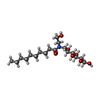
| Nonpolymer details | THE BROMINE ION IS FROM THE COMPOUND AMINO-5-BROMOBENZOTHIAZOLE, THE REMAINDER OF WHICH IS NOT ...THE BROMINE ION IS FROM THE COMPOUND AMINO-5-BROMOBENZO |
|---|
-Experimental details
-Experiment
| Experiment | Method: X-RAY DIFFRACTION |
|---|
- Sample preparation
| Crystal | Density Matthews: 3.94 Å3/Da / Density % sol: 68.77 % / Description: flat rectangular plates |
|---|---|
| Crystal grow | Temperature: 277 K / Method: vapor diffusion, sitting drop / pH: 8 / Details: 0.1 M sodium citrate, 0.1 MTris, 34% PEG |
-Data collection
| Diffraction | Mean temperature: 100 K |
|---|---|
| Diffraction source | Source: SYNCHROTRON / Site: ESRF  / Beamline: ID23-1 / Wavelength: 0.918401 Å / Beamline: ID23-1 / Wavelength: 0.918401 Å |
| Detector | Type: DECTRIS PILATUS 6M-F / Detector: PIXEL / Date: Dec 6, 2013 |
| Radiation | Monochromator: Si(111) crystal / Protocol: SINGLE WAVELENGTH / Monochromatic (M) / Laue (L): M / Scattering type: x-ray |
| Radiation wavelength | Wavelength: 0.918401 Å / Relative weight: 1 |
| Reflection | Resolution: 2.9→45.41 Å / Num. obs: 23548 / % possible obs: 98.7 % / Observed criterion σ(F): 0 / Observed criterion σ(I): 0 / Redundancy: 9.9 % / Rmerge(I) obs: 0.155 / Net I/σ(I): 12.8 |
| Reflection shell | Resolution: 2.9→3.09 Å / Redundancy: 9.3 % / Rmerge(I) obs: 0.581 / Net I/σ(I) obs: 3.7 / % possible all: 92.5 |
- Processing
| Software | Name: BUSTER / Version: 2.10.0 / Classification: refinement | |||||||||||||||||||||||||||||||||||||||||||||||||||||||||||||||||||||||||||||||||||||||||||||||||||||||||||||||||||||||||||||
|---|---|---|---|---|---|---|---|---|---|---|---|---|---|---|---|---|---|---|---|---|---|---|---|---|---|---|---|---|---|---|---|---|---|---|---|---|---|---|---|---|---|---|---|---|---|---|---|---|---|---|---|---|---|---|---|---|---|---|---|---|---|---|---|---|---|---|---|---|---|---|---|---|---|---|---|---|---|---|---|---|---|---|---|---|---|---|---|---|---|---|---|---|---|---|---|---|---|---|---|---|---|---|---|---|---|---|---|---|---|---|---|---|---|---|---|---|---|---|---|---|---|---|---|---|---|---|
| Refinement | Method to determine structure: MOLECULAR REPLACEMENT / Resolution: 2.91→32.73 Å / Cor.coef. Fo:Fc: 0.8874 / Cor.coef. Fo:Fc free: 0.8915 / SU R Cruickshank DPI: 0.262 / Cross valid method: THROUGHOUT / σ(F): 0 / SU R Blow DPI: 0.283 / SU Rfree Blow DPI: 0.217 / SU Rfree Cruickshank DPI: 0.212
| |||||||||||||||||||||||||||||||||||||||||||||||||||||||||||||||||||||||||||||||||||||||||||||||||||||||||||||||||||||||||||||
| Displacement parameters | Biso mean: 62.58 Å2
| |||||||||||||||||||||||||||||||||||||||||||||||||||||||||||||||||||||||||||||||||||||||||||||||||||||||||||||||||||||||||||||
| Refine analyze | Luzzati coordinate error obs: 0.287 Å | |||||||||||||||||||||||||||||||||||||||||||||||||||||||||||||||||||||||||||||||||||||||||||||||||||||||||||||||||||||||||||||
| Refinement step | Cycle: 1 / Resolution: 2.91→32.73 Å
| |||||||||||||||||||||||||||||||||||||||||||||||||||||||||||||||||||||||||||||||||||||||||||||||||||||||||||||||||||||||||||||
| Refine LS restraints |
| |||||||||||||||||||||||||||||||||||||||||||||||||||||||||||||||||||||||||||||||||||||||||||||||||||||||||||||||||||||||||||||
| LS refinement shell | Resolution: 2.91→3.04 Å / Total num. of bins used: 12
| |||||||||||||||||||||||||||||||||||||||||||||||||||||||||||||||||||||||||||||||||||||||||||||||||||||||||||||||||||||||||||||
| Refinement TLS params. | Method: refined / Refine-ID: X-RAY DIFFRACTION
| |||||||||||||||||||||||||||||||||||||||||||||||||||||||||||||||||||||||||||||||||||||||||||||||||||||||||||||||||||||||||||||
| Refinement TLS group |
|
