 Movie
Movie Controller

[English] 日本語
 Yorodumi
- PDB-4p7n: Structure of Escherichia coli PgaB C-terminal domain in complex w... -
+ Open data

- Basic information
| Entry | Database: PDB / ID: 4p7n | ||||||
|---|---|---|---|---|---|---|---|
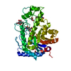
| Title | Structure of Escherichia coli PgaB C-terminal domain in complex with glucosamine | ||||||
 Components Components | Poly-beta-1,6-N-acetyl-D-glucosamine N-deacetylase | ||||||
 Keywords Keywords | HYDROLASE / beta alpha barrel / carbohydrate binding / glycosyl hydrolase fold / complex | ||||||
| Function / homology |  Function and homology information Function and homology informationmacromolecule deacylation / cell adhesion involved in biofilm formation / hydrolase activity, acting on carbon-nitrogen (but not peptide) bonds / single-species biofilm formation / Hydrolases; Acting on carbon-nitrogen bonds, other than peptide bonds; In linear amides / hydrolase activity, hydrolyzing O-glycosyl compounds / cell outer membrane / carbohydrate metabolic process / hydrolase activity Similarity search - Function | ||||||
| Biological species |  | ||||||
| Method |  X-RAY DIFFRACTION / SYNCHROTRON / MOLECULAR REPLACEMENT / molecular replacement / Resolution: 1.89 Å X-RAY DIFFRACTION / SYNCHROTRON / MOLECULAR REPLACEMENT / molecular replacement / Resolution: 1.89 Å | ||||||
 Authors Authors | Little, D.J. / Li, G. / Ing, C. / DiFrancesco, B. / Bamford, N.C. / Robinson, H. / Nitz, M. / Pomes, R. / Howell, P.L. | ||||||
| Funding support |  Canada, 1items Canada, 1items
| ||||||
 Citation Citation | Journal: Proc.Natl.Acad.Sci.USA / Year: 2014 Title: Modification and periplasmic translocation of the biofilm exopolysaccharide poly-beta-1,6-N-acetyl-D-glucosamine. Authors: Little, D.J. / Li, G. / Ing, C. / DiFrancesco, B.R. / Bamford, N.C. / Robinson, H. / Nitz, M. / Pomes, R. / Howell, P.L. | ||||||
| History |
|
- Structure visualization
| Structure viewer | Molecule: MolmilJmol/JSmol |
|---|
- Downloads & links
-Download
| PDBx/mmCIF format | 4p7n.cif.gz | 173.4 KB | Display | PDBx/mmCIF format |
|---|---|---|---|---|
| PDB format | pdb4p7n.ent.gz | 134.2 KB | Display | PDB format |
| PDBx/mmJSON format | 4p7n.json.gz | Tree view | PDBx/mmJSON format | |
| Others |  Other downloads Other downloads |
-Validation report
| Arichive directory | https://data.pdbj.org/pub/pdb/validation_reports/p7/4p7nftp://data.pdbj.org/pub/pdb/validation_reports/p7/4p7n | HTTPS FTP |
|---|
-Related structure data
| Related structure data | 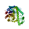 4p7lSC 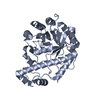 4p7oC  4p7qC 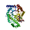 4p7rC S: Starting model for refinement C: citing same article ( |
|---|---|
| Similar structure data |


-Links
 PDBj
PDBj


- Assembly
| Deposited unit | 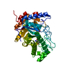
| ||||||||
|---|---|---|---|---|---|---|---|---|---|
| 1 |
| ||||||||
| Unit cell |
| ||||||||
| Details | biological unit is the same as asym. |
-Components
| #1: Protein | Mass: 42441.867 Da / Num. of mol.: 1 / Fragment: UNP residues 310-672 Source method: isolated from a genetically manipulated source Source: (gene. exp.) References: UniProt: P75906, Hydrolases; Acting on carbon-nitrogen bonds, other than peptide bonds; In linear amides | ||
|---|---|---|---|
| #2: Sugar |   Type: D-saccharide, beta linking / Mass: 179.171 Da / Num. of mol.: 3 / Source method: obtained synthetically / Formula: C6H13NO5 Type: D-saccharide, beta linking / Mass: 179.171 Da / Num. of mol.: 3 / Source method: obtained synthetically / Formula: C6H13NO5#3: Water | ChemComp-HOH / |  Mass: 18.015 Da / Num. of mol.: 388 / Source method: isolated from a natural source / Formula: H2O Mass: 18.015 Da / Num. of mol.: 388 / Source method: isolated from a natural source / Formula: H2O |
-Experimental details
-Experiment
| Experiment | Method: X-RAY DIFFRACTION / Number of used crystals: 1 |
|---|
- Sample preparation
| Crystal | Density Matthews: 2.26 Å3/Da / Density % sol: 45.57 % |
|---|---|
| Crystal grow | Temperature: 293 K / Method: vapor diffusion, hanging drop / pH: 6.5 / Details: 18% PEG3350, 0.1 M Bis-Tris |
-Data collection
| Diffraction | Mean temperature: 100 K |
|---|---|
| Diffraction source | Source: SYNCHROTRON / Site: NSLS  / Beamline: X29A / Wavelength: 1.08 Å / Beamline: X29A / Wavelength: 1.08 Å |
| Detector | Type: ADSC QUANTUM 315r / Detector: CCD / Date: Mar 26, 2012 |
| Radiation | Monochromator: Rosenbaum-Rock double crystal sagittal focusing Protocol: SINGLE WAVELENGTH / Monochromatic (M) / Laue (L): M / Scattering type: x-ray |
| Radiation wavelength | Wavelength: 1.08 Å / Relative weight: 1 |
| Reflection | Resolution: 1.89→50 Å / Num. obs: 31672 / % possible obs: 100 % / Observed criterion σ(I): -3 / Redundancy: 14 % / Biso Wilson estimate: 21.71 Å2 / Rmerge(I) obs: 0.138 / Net I/σ(I): 19.9 |
| Reflection shell | Resolution: 1.89→1.96 Å / Redundancy: 13.7 % / Rmerge(I) obs: 0.54 / Mean I/σ(I) obs: 5 / % possible all: 99.9 |
-Phasing
| Phasing | Method: molecular replacement |
|---|
- Processing
| Software |
| |||||||||||||||||||||||||||||||||||||||||||||||||||||||||||||||||||||||||||||||||||||||||||||||||||||||||||||||||||||||||||||||||||||||||||||||||||||||||||||||||||||||||||||||||||||||||||||||||||||||||||||||||||||||||||||||||||||||||||||||||||||||||||||||||||||||||||||||||||
|---|---|---|---|---|---|---|---|---|---|---|---|---|---|---|---|---|---|---|---|---|---|---|---|---|---|---|---|---|---|---|---|---|---|---|---|---|---|---|---|---|---|---|---|---|---|---|---|---|---|---|---|---|---|---|---|---|---|---|---|---|---|---|---|---|---|---|---|---|---|---|---|---|---|---|---|---|---|---|---|---|---|---|---|---|---|---|---|---|---|---|---|---|---|---|---|---|---|---|---|---|---|---|---|---|---|---|---|---|---|---|---|---|---|---|---|---|---|---|---|---|---|---|---|---|---|---|---|---|---|---|---|---|---|---|---|---|---|---|---|---|---|---|---|---|---|---|---|---|---|---|---|---|---|---|---|---|---|---|---|---|---|---|---|---|---|---|---|---|---|---|---|---|---|---|---|---|---|---|---|---|---|---|---|---|---|---|---|---|---|---|---|---|---|---|---|---|---|---|---|---|---|---|---|---|---|---|---|---|---|---|---|---|---|---|---|---|---|---|---|---|---|---|---|---|---|---|---|---|---|---|---|---|---|---|---|---|---|---|---|---|---|---|---|---|---|---|---|---|---|---|---|---|---|---|---|---|---|---|---|---|---|---|---|---|---|---|---|---|---|---|---|---|---|---|---|---|
| Refinement | Method to determine structure: MOLECULAR REPLACEMENT Starting model: PDB entry 4P7L Resolution: 1.89→39.416 Å / FOM work R set: 0.8908 / SU ML: 0.19 / Cross valid method: FREE R-VALUE / σ(F): 1.36 / Phase error: 17.82 / Stereochemistry target values: ML
| |||||||||||||||||||||||||||||||||||||||||||||||||||||||||||||||||||||||||||||||||||||||||||||||||||||||||||||||||||||||||||||||||||||||||||||||||||||||||||||||||||||||||||||||||||||||||||||||||||||||||||||||||||||||||||||||||||||||||||||||||||||||||||||||||||||||||||||||||||
| Solvent computation | Shrinkage radii: 0.9 Å / VDW probe radii: 1.11 Å / Solvent model: FLAT BULK SOLVENT MODEL | |||||||||||||||||||||||||||||||||||||||||||||||||||||||||||||||||||||||||||||||||||||||||||||||||||||||||||||||||||||||||||||||||||||||||||||||||||||||||||||||||||||||||||||||||||||||||||||||||||||||||||||||||||||||||||||||||||||||||||||||||||||||||||||||||||||||||||||||||||
| Displacement parameters | Biso max: 119.18 Å2 / Biso mean: 26.48 Å2 / Biso min: 9.36 Å2 | |||||||||||||||||||||||||||||||||||||||||||||||||||||||||||||||||||||||||||||||||||||||||||||||||||||||||||||||||||||||||||||||||||||||||||||||||||||||||||||||||||||||||||||||||||||||||||||||||||||||||||||||||||||||||||||||||||||||||||||||||||||||||||||||||||||||||||||||||||
| Refinement step | Cycle: final / Resolution: 1.89→39.416 Å
| |||||||||||||||||||||||||||||||||||||||||||||||||||||||||||||||||||||||||||||||||||||||||||||||||||||||||||||||||||||||||||||||||||||||||||||||||||||||||||||||||||||||||||||||||||||||||||||||||||||||||||||||||||||||||||||||||||||||||||||||||||||||||||||||||||||||||||||||||||
| Refine LS restraints |
| |||||||||||||||||||||||||||||||||||||||||||||||||||||||||||||||||||||||||||||||||||||||||||||||||||||||||||||||||||||||||||||||||||||||||||||||||||||||||||||||||||||||||||||||||||||||||||||||||||||||||||||||||||||||||||||||||||||||||||||||||||||||||||||||||||||||||||||||||||
| LS refinement shell | Refine-ID: X-RAY DIFFRACTION / Total num. of bins used: 27
| |||||||||||||||||||||||||||||||||||||||||||||||||||||||||||||||||||||||||||||||||||||||||||||||||||||||||||||||||||||||||||||||||||||||||||||||||||||||||||||||||||||||||||||||||||||||||||||||||||||||||||||||||||||||||||||||||||||||||||||||||||||||||||||||||||||||||||||||||||
| Refinement TLS params. | Method: refined / Refine-ID: X-RAY DIFFRACTION
| |||||||||||||||||||||||||||||||||||||||||||||||||||||||||||||||||||||||||||||||||||||||||||||||||||||||||||||||||||||||||||||||||||||||||||||||||||||||||||||||||||||||||||||||||||||||||||||||||||||||||||||||||||||||||||||||||||||||||||||||||||||||||||||||||||||||||||||||||||
| Refinement TLS group |
|
