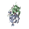
 Movie
Movie Controller


+ Open data

- Basic information
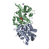
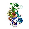
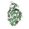
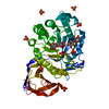
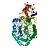
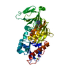
| Entry | Database: PDB / ID: 4p6c | ||||||
|---|---|---|---|---|---|---|---|
| Title | Structure of ribB complexed with inhibitor 4PEH | ||||||
 Components Components | 3,4-dihydroxy-2-butanone 4-phosphate synthase | ||||||
 Keywords Keywords | LYASE/LYASE INHIBITOR / ribB / DHBPS / inhibitor / riboflavin / complex / 4PEH / LYASE-LYASE INHIBITOR complex | ||||||
| Function / homology |  Function and homology information Function and homology information3,4-dihydroxy-2-butanone-4-phosphate synthase / 3,4-dihydroxy-2-butanone-4-phosphate synthase activity / riboflavin biosynthetic process / manganese ion binding / magnesium ion binding / cytosol Similarity search - Function | ||||||
| Biological species |  Vibrio cholerae serotype O1 (bacteria) Vibrio cholerae serotype O1 (bacteria) | ||||||
| Method |  X-RAY DIFFRACTION / MOLECULAR REPLACEMENT / Resolution: 1.86 Å X-RAY DIFFRACTION / MOLECULAR REPLACEMENT / Resolution: 1.86 Å | ||||||
 Authors Authors | Islam, Z. / Kumar, A. / Singh, S. / Salmon, L. / Karthikeyan, S. | ||||||
| Funding support |  India, 1items India, 1items
| ||||||
 Citation Citation | Journal: J.Biol.Chem. / Year: 2015 Title: Structural Basis for Competitive Inhibition of 3,4-Dihydroxy-2-butanone-4-phosphate Synthase from Vibrio cholerae. Authors: Islam, Z. / Kumar, A. / Singh, S. / Salmon, L. / Karthikeyan, S. | ||||||
| History |
|
- Structure visualization
| Structure viewer | Molecule: MolmilJmol/JSmol |
|---|
- Downloads & links
-Download
| PDBx/mmCIF format | 4p6c.cif.gz | 102.8 KB | Display | PDBx/mmCIF format |
|---|---|---|---|---|
| PDB format | pdb4p6c.ent.gz | 77.2 KB | Display | PDB format |
| PDBx/mmJSON format | 4p6c.json.gz | Tree view | PDBx/mmJSON format | |
| Others |  Other downloads Other downloads |
-Validation report
| Summary document | 4p6c_validation.pdf.gz | 451.3 KB | Display | wwPDB validaton report |
|---|---|---|---|---|
| Full document | 4p6c_full_validation.pdf.gz | 454.7 KB | Display | |
| Data in XML | 4p6c_validation.xml.gz | 21.1 KB | Display | |
| Data in CIF | 4p6c_validation.cif.gz | 30.7 KB | Display | |
| Arichive directory | https://data.pdbj.org/pub/pdb/validation_reports/p6/4p6cftp://data.pdbj.org/pub/pdb/validation_reports/p6/4p6c | HTTPS FTP |
-Related structure data
| Related structure data |  4p6dC  4p6pC  4p77C 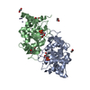 4p8eC  4p8jC 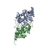 1g57S C: citing same article ( S: Starting model for refinement |
|---|---|
| Similar structure data |

-Links
 PDBj
PDBj- Assembly
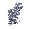
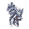
| Deposited unit | 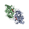
| ||||||||
|---|---|---|---|---|---|---|---|---|---|
| 1 |
| ||||||||
| Unit cell |
|
-Components
| #1: Protein | Mass: 25761.328 Da / Num. of mol.: 2 Source method: isolated from a genetically manipulated source Source: (gene. exp.) Vibrio cholerae serotype O1 (bacteria) / Strain: ATCC 39315 / El Tor Inaba N16961 / Gene: ribB, VC_A1060 / Plasmid: pET28c / Production host: References: UniProt: Q9KKP2, 3,4-dihydroxy-2-butanone-4-phosphate synthase #2: Chemical | 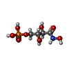  Mass: 231.098 Da / Num. of mol.: 2 / Source method: obtained synthetically / Formula: C4H10NO8P Mass: 231.098 Da / Num. of mol.: 2 / Source method: obtained synthetically / Formula: C4H10NO8P#3: Water | ChemComp-HOH / |  Mass: 18.015 Da / Num. of mol.: 344 / Source method: isolated from a natural source / Formula: H2O Mass: 18.015 Da / Num. of mol.: 344 / Source method: isolated from a natural source / Formula: H2O |
|---|
-Experimental details
-Experiment
| Experiment | Method: X-RAY DIFFRACTION / Number of used crystals: 1 |
|---|
- Sample preparation
| Crystal | Density Matthews: 1.92 Å3/Da / Density % sol: 35.8 % / Description: Rod shape |
|---|---|
| Crystal grow | Temperature: 293 K / Method: vapor diffusion, sitting drop / pH: 9.1 / Details: PEG3350, Na2HPO4 |
-Data collection
| Diffraction | Mean temperature: 100 K |
|---|---|
| Diffraction source | Source: ROTATING ANODE / Type: RIGAKU MICROMAX-007 HF / Wavelength: 1.5418 Å |
| Detector | Type: MAR scanner 345 mm plate / Detector: IMAGE PLATE / Date: Jun 24, 2010 / Details: Mirror |
| Radiation | Monochromator: Mirror / Protocol: SINGLE WAVELENGTH / Monochromatic (M) / Laue (L): M / Scattering type: x-ray |
| Radiation wavelength | Wavelength: 1.5418 Å / Relative weight: 1 |
| Reflection | Resolution: 1.86→20 Å / Num. obs: 32615 / % possible obs: 99.3 % / Observed criterion σ(I): -3 / Redundancy: 7.7 % / Biso Wilson estimate: 14.62 Å2 / Rmerge(I) obs: 0.105 / Net I/σ(I): 15.7 |
| Reflection shell | Resolution: 1.86→1.97 Å / Redundancy: 7.6 % / Rmerge(I) obs: 0.361 / Mean I/σ(I) obs: 5.5 / % possible all: 97.7 |
- Processing
| Software |
| |||||||||||||||||||||||||||||||||||||||||||||||||||||||||||||||||||||||||||||||||||||||||||
|---|---|---|---|---|---|---|---|---|---|---|---|---|---|---|---|---|---|---|---|---|---|---|---|---|---|---|---|---|---|---|---|---|---|---|---|---|---|---|---|---|---|---|---|---|---|---|---|---|---|---|---|---|---|---|---|---|---|---|---|---|---|---|---|---|---|---|---|---|---|---|---|---|---|---|---|---|---|---|---|---|---|---|---|---|---|---|---|---|---|---|---|---|
| Refinement | Method to determine structure: MOLECULAR REPLACEMENT Starting model: 1G57 Resolution: 1.86→19.62 Å / SU ML: 0.17 / Cross valid method: FREE R-VALUE / σ(F): 1.99 / Phase error: 18.19 / Stereochemistry target values: ML
| |||||||||||||||||||||||||||||||||||||||||||||||||||||||||||||||||||||||||||||||||||||||||||
| Solvent computation | Shrinkage radii: 0.9 Å / VDW probe radii: 1.11 Å / Solvent model: FLAT BULK SOLVENT MODEL | |||||||||||||||||||||||||||||||||||||||||||||||||||||||||||||||||||||||||||||||||||||||||||
| Displacement parameters | Biso mean: 13.4 Å2 | |||||||||||||||||||||||||||||||||||||||||||||||||||||||||||||||||||||||||||||||||||||||||||
| Refinement step | Cycle: LAST / Resolution: 1.86→19.62 Å
| |||||||||||||||||||||||||||||||||||||||||||||||||||||||||||||||||||||||||||||||||||||||||||
| Refine LS restraints |
| |||||||||||||||||||||||||||||||||||||||||||||||||||||||||||||||||||||||||||||||||||||||||||
| LS refinement shell |
|
