 Movie
Movie Controller

[English] 日本語
 Yorodumi
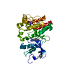
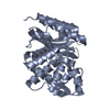
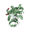
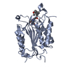
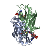
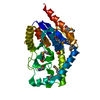
- PDB-4p5d: CRYSTAL STRUCTURE OF RAT DNPH1 (RCL) WITH 6-NAPHTHYL-PURINE-RIBOS... -
+ Open data

- Basic information
| Entry | Database: PDB / ID: 4p5d | ||||||
|---|---|---|---|---|---|---|---|
| Title | CRYSTAL STRUCTURE OF RAT DNPH1 (RCL) WITH 6-NAPHTHYL-PURINE-RIBOSIDE-MONOPHOSPHATE | ||||||
 Components Components | 2'-deoxynucleoside 5'-phosphate N-hydrolase 1 | ||||||
 Keywords Keywords | HYDROLASE / N-GLYCOSIDASE / INHIBITOR | ||||||
| Function / homology |  Function and homology information Function and homology informationPurine catabolism / 5-hydroxymethyl-dUMP N-hydrolase activity / nucleoside salvage / deoxyribonucleoside monophosphate catabolic process / dGMP catabolic process / allantoin metabolic process / Hydrolases; Glycosylases; Hydrolysing N-glycosyl compounds / epithelial cell differentiation / positive regulation of cell growth / protein homodimerization activity ...Purine catabolism / 5-hydroxymethyl-dUMP N-hydrolase activity / nucleoside salvage / deoxyribonucleoside monophosphate catabolic process / dGMP catabolic process / allantoin metabolic process / Hydrolases; Glycosylases; Hydrolysing N-glycosyl compounds / epithelial cell differentiation / positive regulation of cell growth / protein homodimerization activity / identical protein binding / nucleus / cytoplasm Similarity search - Function | ||||||
| Biological species |  | ||||||
| Method |  X-RAY DIFFRACTION / SYNCHROTRON / MOLECULAR REPLACEMENT / Resolution: 2.11 Å X-RAY DIFFRACTION / SYNCHROTRON / MOLECULAR REPLACEMENT / Resolution: 2.11 Å | ||||||
 Authors Authors | Padilla, A. / Labesse, G. / Kaminski, P.A. | ||||||
 Citation Citation | Journal: Eur.J.Med.Chem. / Year: 2014 Title: 6-(Hetero)Arylpurine nucleotides as inhibitors of the oncogenic target DNPH1: Synthesis, structural studies and cytotoxic activities. Authors: Amiable, C. / Paoletti, J. / Haouz, A. / Padilla, A. / Labesse, G. / Kaminski, P.A. / Pochet, S. #1: Journal: Acta Crystallogr. D Biol. Crystallogr. / Year: 2013Title: Structure of the oncoprotein Rcl bound to three nucleotide analogues. Authors: Padilla, A. / Amiable, C. / Pochet, S. / Kaminski, P.A. / Labesse, G. | ||||||
| History |
|
- Structure visualization
| Structure viewer | Molecule: MolmilJmol/JSmol |
|---|
- Downloads & links
-Download
| PDBx/mmCIF format | 4p5d.cif.gz | 138.2 KB | Display | PDBx/mmCIF format |
|---|---|---|---|---|
| PDB format | pdb4p5d.ent.gz | 107.9 KB | Display | PDB format |
| PDBx/mmJSON format | 4p5d.json.gz | Tree view | PDBx/mmJSON format | |
| Others |  Other downloads Other downloads |
-Validation report
| Arichive directory | https://data.pdbj.org/pub/pdb/validation_reports/p5/4p5dftp://data.pdbj.org/pub/pdb/validation_reports/p5/4p5d | HTTPS FTP |
|---|
-Related structure data
| Related structure data |  4p5eC  4fyiS S: Starting model for refinement C: citing same article ( |
|---|---|
| Similar structure data |


-Links
 PDBj
PDBj- Assembly
| Deposited unit | 
| ||||||||
|---|---|---|---|---|---|---|---|---|---|
| 1 | 
| ||||||||
| 2 | 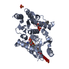
| ||||||||
| Unit cell |
| ||||||||
| Components on special symmetry positions |
|
-Components
| #1: Protein | Mass: 17173.334 Da / Num. of mol.: 2 / Fragment: Residues 11-151 / Mutation: yes Source method: isolated from a genetically manipulated source Source: (gene. exp.)  References: UniProt: O35820, Hydrolases; Glycosylases; Hydrolysing N-glycosyl compounds #2: Chemical |   Mass: 458.361 Da / Num. of mol.: 2 / Source method: obtained synthetically / Formula: C20H19N4O7P Mass: 458.361 Da / Num. of mol.: 2 / Source method: obtained synthetically / Formula: C20H19N4O7P#3: Chemical | ChemComp-SO4 / |   Mass: 96.063 Da / Num. of mol.: 1 / Source method: obtained synthetically / Formula: SO4 Mass: 96.063 Da / Num. of mol.: 1 / Source method: obtained synthetically / Formula: SO4#4: Water | ChemComp-HOH / |  Mass: 18.015 Da / Num. of mol.: 144 / Source method: isolated from a natural source / Formula: H2O Mass: 18.015 Da / Num. of mol.: 144 / Source method: isolated from a natural source / Formula: H2O |
|---|
-Experimental details
-Experiment
| Experiment | Method: X-RAY DIFFRACTION |
|---|
- Sample preparation
| Crystal | Density Matthews: 3.8 Å3/Da / Density % sol: 67.69 % |
|---|---|
| Crystal grow | Temperature: 291 K / Method: vapor diffusion, hanging drop / pH: 8 Details: 1.3 M LITHIUM SULFATE, 1% PEG2K, 20 MM MAGNESIUM SULFATE, 100 MM TRIS, PH 8.0 |
-Data collection
| Diffraction | Mean temperature: 100 K |
|---|---|
| Diffraction source | Source: SYNCHROTRON / Site: ESRF  / Beamline: ID29 / Wavelength: 0.9793 Å / Beamline: ID29 / Wavelength: 0.9793 Å |
| Detector | Type: PSI PILATUS 6M / Detector: PIXEL / Date: Mar 9, 2011 |
| Radiation | Protocol: SINGLE WAVELENGTH / Monochromatic (M) / Laue (L): M / Scattering type: x-ray |
| Radiation wavelength | Wavelength: 0.9793 Å / Relative weight: 1 |
| Reflection | Resolution: 2.109→38.59 Å / Num. obs: 31129 / % possible obs: 99.4 % / Redundancy: 6.5 % / Rmerge(I) obs: 0.066 / Net I/σ(I): 15.9 |
| Reflection shell | Resolution: 2.11→2.22 Å / % possible obs: 99.5 % / Redundancy: 6.9 % / Rmerge(I) obs: 0.54 / Mean I/σ(I) obs: 3.9 |
- Processing
| Software |
| ||||||||||||||||||||||||||||||||||||||||||||||||||||||||||||||||||||||||||||||||||||
|---|---|---|---|---|---|---|---|---|---|---|---|---|---|---|---|---|---|---|---|---|---|---|---|---|---|---|---|---|---|---|---|---|---|---|---|---|---|---|---|---|---|---|---|---|---|---|---|---|---|---|---|---|---|---|---|---|---|---|---|---|---|---|---|---|---|---|---|---|---|---|---|---|---|---|---|---|---|---|---|---|---|---|---|---|---|
| Refinement | Method to determine structure: MOLECULAR REPLACEMENT Starting model: 4FYI Resolution: 2.11→38.165 Å / SU ML: 0.18 / Cross valid method: THROUGHOUT / σ(F): 1.39 / Phase error: 18.62 / Stereochemistry target values: ML
| ||||||||||||||||||||||||||||||||||||||||||||||||||||||||||||||||||||||||||||||||||||
| Solvent computation | Shrinkage radii: 0.9 Å / VDW probe radii: 1.11 Å / Solvent model: FLAT BULK SOLVENT MODEL | ||||||||||||||||||||||||||||||||||||||||||||||||||||||||||||||||||||||||||||||||||||
| Refinement step | Cycle: LAST / Resolution: 2.11→38.165 Å
| ||||||||||||||||||||||||||||||||||||||||||||||||||||||||||||||||||||||||||||||||||||
| Refine LS restraints |
| ||||||||||||||||||||||||||||||||||||||||||||||||||||||||||||||||||||||||||||||||||||
| LS refinement shell |
| ||||||||||||||||||||||||||||||||||||||||||||||||||||||||||||||||||||||||||||||||||||
| Refinement TLS params. | Method: refined / Refine-ID: X-RAY DIFFRACTION
| ||||||||||||||||||||||||||||||||||||||||||||||||||||||||||||||||||||||||||||||||||||
| Refinement TLS group |
|
