 Movie
Movie Controller


+ Open data

- Basic information
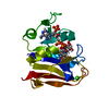
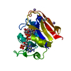
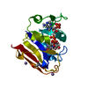
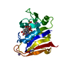
| Entry | Database: PDB / ID: 4p3r | ||||||
|---|---|---|---|---|---|---|---|
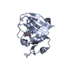
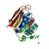
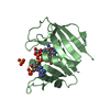
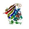
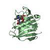
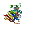
| Title | Cryogenic WT DHFR, time-averaged ensemble | ||||||
 Components Components | Dihydrofolate reductase | ||||||
 Keywords Keywords | OXIDOREDUCTASE / ROSSMANN FOLD | ||||||
| Function / homology | Dihydrofolate Reductase, subunit A / Dihydrofolate Reductase, subunit A / 3-Layer(aba) Sandwich / Alpha Beta / FOLIC ACID / NADP NICOTINAMIDE-ADENINE-DINUCLEOTIDE PHOSPHATE / :  Function and homology information Function and homology information | ||||||
| Biological species |  | ||||||
| Method |  X-RAY DIFFRACTION / SYNCHROTRON / MOLECULAR REPLACEMENT / Resolution: 1.15 Å X-RAY DIFFRACTION / SYNCHROTRON / MOLECULAR REPLACEMENT / Resolution: 1.15 Å | ||||||
 Authors Authors | Keedy, D.A. / van den Bedem, H. / Fraser, J.S. | ||||||
 Citation Citation | Journal: Structure / Year: 2014 Title: Crystal Cryocooling Distorts Conformational Heterogeneity in a Model Michaelis Complex of DHFR. Authors: Keedy, D.A. / van den Bedem, H. / Sivak, D.A. / Petsko, G.A. / Ringe, D. / Wilson, M.A. / Fraser, J.S. | ||||||
| History |
|
- Structure visualization
| Structure viewer | Molecule: MolmilJmol/JSmol |
|---|
- Downloads & links
-Download
| PDBx/mmCIF format | 4p3r.cif.gz | 28.8 MB | Display | PDBx/mmCIF format |
|---|---|---|---|---|
| PDB format | pdb4p3r.ent.gz | 24.1 MB | Display | PDB format |
| PDBx/mmJSON format | 4p3r.json.gz | Tree view | PDBx/mmJSON format | |
| Others |  Other downloads Other downloads |
-Validation report
| Arichive directory | https://data.pdbj.org/pub/pdb/validation_reports/p3/4p3rftp://data.pdbj.org/pub/pdb/validation_reports/p3/4p3r | HTTPS FTP |
|---|
-Related structure data


-Links
 PDBj
PDBj
- Assembly
| Deposited unit | 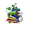
| ||||||||||
|---|---|---|---|---|---|---|---|---|---|---|---|
| 1 |
| ||||||||||
| Unit cell |
| ||||||||||
| Number of models | 250 |
-Components
| #1: Protein | Mass: 18019.340 Da / Num. of mol.: 1 Source method: isolated from a genetically manipulated source Source: (gene. exp.) |
|---|---|
| #2: Chemical | ChemComp-FOL /   Mass: 441.397 Da / Num. of mol.: 1 / Source method: obtained synthetically / Formula: C19H19N7O6 Mass: 441.397 Da / Num. of mol.: 1 / Source method: obtained synthetically / Formula: C19H19N7O6 |
| #3: Chemical | ChemComp-NAP /   Mass: 743.405 Da / Num. of mol.: 1 / Source method: obtained synthetically / Formula: C21H28N7O17P3 Mass: 743.405 Da / Num. of mol.: 1 / Source method: obtained synthetically / Formula: C21H28N7O17P3 |
| #4: Water | ChemComp-HOH /  Mass: 18.015 Da / Num. of mol.: 200 / Source method: isolated from a natural source / Formula: H2O Mass: 18.015 Da / Num. of mol.: 200 / Source method: isolated from a natural source / Formula: H2O |
-Experimental details
-Experiment
| Experiment | Method: X-RAY DIFFRACTION / Number of used crystals: 1 |
|---|
- Sample preparation
| Crystal | Density Matthews: 2.08 Å3/Da / Density % sol: 40.83 % |
|---|---|
| Crystal grow | Temperature: 293 K / Method: vapor diffusion, hanging drop / pH: 7.5 Details: 100MM HEPES PH 7.5, 21% PEG8000, 200MM MGCL2, VAPOR DIFFUSION, HANGING DROP, TEMPERATURE 293K |
-Data collection
| Diffraction | Mean temperature: 100 K |
|---|---|
| Diffraction source | Source: SYNCHROTRON / Site: ALS  / Beamline: 8.3.1 / Wavelength: 1.115869 Å / Beamline: 8.3.1 / Wavelength: 1.115869 Å |
| Detector | Type: ADSC QUANTUM 315 / Detector: CCD / Date: Oct 25, 2011 |
| Radiation | Monochromator: KOHZU DUAL DOUBLE CRYSTAL MONOCHROMATOR / Protocol: SINGLE WAVELENGTH / Monochromatic (M) / Laue (L): M / Scattering type: x-ray |
| Radiation wavelength | Wavelength: 1.115869 Å / Relative weight: 1 |
| Reflection | Resolution: 1.15→50 Å / Num. obs: 51212 / % possible obs: 94.4 % / Observed criterion σ(I): 0 / Redundancy: 4.9 % / Net I/σ(I): 22.6 |
| Reflection shell | Resolution: 1.15→1.19 Å / % possible all: 64.2 |
- Processing
| Software |
| ||||||||||||||||||||||||||||||||||||||||||||||||||||||||||||||||||||||||||||||||||||||||||||||||||||||||||||||||||||||||||||||||||||||||||||
|---|---|---|---|---|---|---|---|---|---|---|---|---|---|---|---|---|---|---|---|---|---|---|---|---|---|---|---|---|---|---|---|---|---|---|---|---|---|---|---|---|---|---|---|---|---|---|---|---|---|---|---|---|---|---|---|---|---|---|---|---|---|---|---|---|---|---|---|---|---|---|---|---|---|---|---|---|---|---|---|---|---|---|---|---|---|---|---|---|---|---|---|---|---|---|---|---|---|---|---|---|---|---|---|---|---|---|---|---|---|---|---|---|---|---|---|---|---|---|---|---|---|---|---|---|---|---|---|---|---|---|---|---|---|---|---|---|---|---|---|---|---|
| Refinement | Method to determine structure: MOLECULAR REPLACEMENT / Resolution: 1.15→40.774 Å / SU ML: 0.04 / Cross valid method: FREE R-VALUE / σ(F): 1.34 / Phase error: 11.37
| ||||||||||||||||||||||||||||||||||||||||||||||||||||||||||||||||||||||||||||||||||||||||||||||||||||||||||||||||||||||||||||||||||||||||||||
| Solvent computation | Shrinkage radii: 0.6 Å / VDW probe radii: 0.8 Å / Solvent model: FLAT BULK SOLVENT MODEL | ||||||||||||||||||||||||||||||||||||||||||||||||||||||||||||||||||||||||||||||||||||||||||||||||||||||||||||||||||||||||||||||||||||||||||||
| Refinement step | Cycle: LAST / Resolution: 1.15→40.774 Å
| ||||||||||||||||||||||||||||||||||||||||||||||||||||||||||||||||||||||||||||||||||||||||||||||||||||||||||||||||||||||||||||||||||||||||||||
| Refine LS restraints |
| ||||||||||||||||||||||||||||||||||||||||||||||||||||||||||||||||||||||||||||||||||||||||||||||||||||||||||||||||||||||||||||||||||||||||||||
| LS refinement shell |
|
