 Movie
Movie Controller

[English] 日本語
 Yorodumi
- PDB-4or6: Structure of Influenza B PB2 cap-binding domain with Q325F mutati... -
+ Open data

- Basic information
| Entry | Database: PDB / ID: 4or6 | ||||||
|---|---|---|---|---|---|---|---|
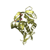
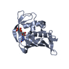
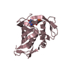
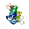
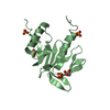
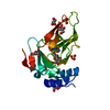
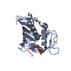
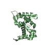
| Title | Structure of Influenza B PB2 cap-binding domain with Q325F mutation complex with GDP | ||||||
 Components Components | Polymerase basic protein 2 | ||||||
 Keywords Keywords | VIRAL PROTEIN / cap binding | ||||||
| Function / homology | : / Influenza RNA polymerase PB2 CAP binding domain / host cell mitochondrion / GUANOSINE-5'-DIPHOSPHATE / Polymerase basic protein 2 Function and homology information Function and homology information | ||||||
| Biological species |  influenza B virus influenza B virus | ||||||
| Method |  X-RAY DIFFRACTION / SYNCHROTRON / MOLECULAR REPLACEMENT / Resolution: 2.293 Å X-RAY DIFFRACTION / SYNCHROTRON / MOLECULAR REPLACEMENT / Resolution: 2.293 Å | ||||||
 Authors Authors | Liu, Y. / Fan, J. / Zheng, X. | ||||||
 Citation Citation | Journal: J.Biol.Chem. / Year: 2015 Title: The crystal structure of the PB2 cap-binding domain of influenza B virus reveals a novel cap recognition mechanism. Authors: Liu, Y. / Yang, Y. / Fan, J. / He, R. / Luo, M. / Zheng, X. | ||||||
| History |
|
- Structure visualization
| Structure viewer | Molecule: MolmilJmol/JSmol |
|---|
- Downloads & links
-Download
| PDBx/mmCIF format | 4or6.cif.gz | 84.7 KB | Display | PDBx/mmCIF format |
|---|---|---|---|---|
| PDB format | pdb4or6.ent.gz | 62.7 KB | Display | PDB format |
| PDBx/mmJSON format | 4or6.json.gz | Tree view | PDBx/mmJSON format | |
| Others |  Other downloads Other downloads |
-Validation report
| Arichive directory | https://data.pdbj.org/pub/pdb/validation_reports/or/4or6ftp://data.pdbj.org/pub/pdb/validation_reports/or/4or6 | HTTPS FTP |
|---|
-Related structure data
| Related structure data |  4or4SC  4q46C S: Starting model for refinement C: citing same article ( |
|---|---|
| Similar structure data |


-Links
 PDBj
PDBj- Assembly
| Deposited unit | 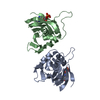
| ||||||||
|---|---|---|---|---|---|---|---|---|---|
| 1 | 
| ||||||||
| 2 | 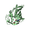
| ||||||||
| Unit cell |
|
-Components
| #1: Protein | Mass: 19629.664 Da / Num. of mol.: 2 / Fragment: cap-binding domain / Mutation: Q325F Source method: isolated from a genetically manipulated source Source: (gene. exp.) influenza B virus / Strain: B/Jiangxi/BV/2006 / Plasmid: pET28a / Production host:  #2: Chemical |   Type: RNA linking / Mass: 443.201 Da / Num. of mol.: 2 / Source method: obtained synthetically / Formula: C10H15N5O11P2 / Comment: GDP, energy-carrying molecule*YM Type: RNA linking / Mass: 443.201 Da / Num. of mol.: 2 / Source method: obtained synthetically / Formula: C10H15N5O11P2 / Comment: GDP, energy-carrying molecule*YM#3: Water | ChemComp-HOH / |  Mass: 18.015 Da / Num. of mol.: 158 / Source method: isolated from a natural source / Formula: H2O Mass: 18.015 Da / Num. of mol.: 158 / Source method: isolated from a natural source / Formula: H2OSequence details | THIS SEQUENCE IS FROM INFLUENZA B VIRUS (B/JIANGXI/BV/2006) WHICH WAS NOT SUBMITTED IN UNP. ...THIS SEQUENCE IS FROM INFLUENZA B VIRUS (B/JIANGXI/BV/2006) WHICH WAS NOT SUBMITTED IN UNP. RESIUDES 316-319 ARE THE EXPRESSION | |
|---|
-Experimental details
-Experiment
| Experiment | Method: X-RAY DIFFRACTION / Number of used crystals: 1 |
|---|
- Sample preparation
| Crystal | Density Matthews: 2.36 Å3/Da / Density % sol: 47.91 % |
|---|---|
| Crystal grow | Temperature: 293 K / Method: vapor diffusion, sitting drop / pH: 7 Details: 20% PEG 3350, 0.15M DL-Malic acid, pH 7.0, VAPOR DIFFUSION, SITTING DROP, temperature 293K |
-Data collection
| Diffraction | Mean temperature: 100 K |
|---|---|
| Diffraction source | Source: SYNCHROTRON / Site: SSRF  / Beamline: BL17U / Wavelength: 1 Å / Beamline: BL17U / Wavelength: 1 Å |
| Detector | Type: ADSC QUANTUM 315r / Detector: CCD / Date: Dec 12, 2013 |
| Radiation | Monochromator: Crystallogic / Protocol: SINGLE WAVELENGTH / Monochromatic (M) / Laue (L): M / Scattering type: x-ray |
| Radiation wavelength | Wavelength: 1 Å / Relative weight: 1 |
| Reflection | Resolution: 2.29→50 Å / Num. all: 17196 / Num. obs: 17179 / % possible obs: 99.9 % / Observed criterion σ(I): 3.8 / Redundancy: 9.3 % / Biso Wilson estimate: 23.52 Å2 |
| Reflection shell | Resolution: 2.29→2.36 Å / % possible all: 99.9 |
- Processing
| Software |
| ||||||||||||||||||||||||||||||||||||||||||
|---|---|---|---|---|---|---|---|---|---|---|---|---|---|---|---|---|---|---|---|---|---|---|---|---|---|---|---|---|---|---|---|---|---|---|---|---|---|---|---|---|---|---|---|
| Refinement | Method to determine structure: MOLECULAR REPLACEMENT Starting model: 4OR4 Resolution: 2.293→30.134 Å / Cor.coef. Fo:Fc: 0.954 / Cor.coef. Fo:Fc free: 0.895 / SU ML: 0.27 / σ(F): 0.72 / Phase error: 22.94 / Stereochemistry target values: ML / Details: HYDROGENS HAVE BEEN ADDED IN THE RIDING POSITIONS
| ||||||||||||||||||||||||||||||||||||||||||
| Solvent computation | Shrinkage radii: 0.9 Å / VDW probe radii: 1.11 Å / Solvent model: FLAT BULK SOLVENT MODEL | ||||||||||||||||||||||||||||||||||||||||||
| Displacement parameters | Biso mean: 22.764 Å2
| ||||||||||||||||||||||||||||||||||||||||||
| Refinement step | Cycle: LAST / Resolution: 2.293→30.134 Å
| ||||||||||||||||||||||||||||||||||||||||||
| Refine LS restraints |
| ||||||||||||||||||||||||||||||||||||||||||
| LS refinement shell | Refine-ID: X-RAY DIFFRACTION / Total num. of bins used: 6
|
