| Entry | Database: PDB / ID: 4ooa
|
|---|
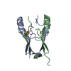
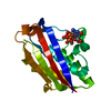
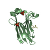
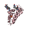
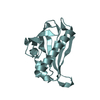
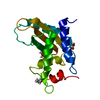
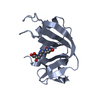
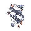
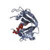
| Title | CRYSTAL STRUCTURE of NAF1 (MINER1): H114C THE REDOX-ACTIVE 2FE-2S PROTEIN |
|---|
 Components Components | CDGSH iron-sulfur domain-containing protein 2 |
|---|
 Keywords Keywords | METAL BINDING PROTEIN / MEMBRANE BOUND / THIAZOLIDINEDIONE / OXIDATIVE STRESS |
|---|
| Function / homology |  Function and homology information Function and homology information
perinuclear endoplasmic reticulum / autophagy of mitochondrion / regulation of autophagy / 2 iron, 2 sulfur cluster binding / mitochondrial outer membrane / endoplasmic reticulum membrane / endoplasmic reticulum / protein homodimerization activity / protein-containing complex / RNA binding ...perinuclear endoplasmic reticulum / autophagy of mitochondrion / regulation of autophagy / 2 iron, 2 sulfur cluster binding / mitochondrial outer membrane / endoplasmic reticulum membrane / endoplasmic reticulum / protein homodimerization activity / protein-containing complex / RNA binding / membrane / metal ion bindingSimilarity search - Function CDGSH iron-sulfur domain, mitoNEET-type / Iron sulphur domain-containing, mitoNEET, N-terminal / Iron-containing outer mitochondrial membrane protein N-terminus / CDGSH iron-sulfur domain-containing protein 1/2 / Iron-binding zinc finger CDGSH type / Iron-binding zinc finger, CDGSH type / MitoNEET, CDGSH iron-sulfur domain / CDGSH-type zinc finger. Function unknown. / Ribosomal Protein L9; domain 1 / 3-Layer(aba) Sandwich / Alpha BetaSimilarity search - Domain/homology |
|---|
| Biological species |  Homo sapiens (human) Homo sapiens (human) |
|---|
| Method |  X-RAY DIFFRACTION / SYNCHROTRON / MOLECULAR REPLACEMENT / Resolution: 1.58 Å X-RAY DIFFRACTION / SYNCHROTRON / MOLECULAR REPLACEMENT / Resolution: 1.58 Å |
|---|
 Authors Authors | Tamir, S. / Eisenberg-Domovich, Y. / Conlan, A.R. / Stofleth, J.T. / Lipper, C.H. / Paddock, M.L. / Mittler, R. / Jennings, P.A. / Livnah, O. / Nechushtai, R. |
|---|
 Citation Citation | Journal: Acta Crystallogr.,Sect.D / Year: 2014
Title: A point mutation in the [2Fe-2S] cluster binding region of the NAF-1 protein (H114C) dramatically hinders the cluster donor properties.
Authors: Tamir, S. / Eisenberg-Domovich, Y. / Conlan, A.R. / Stofleth, J.T. / Lipper, C.H. / Paddock, M.L. / Mittler, R. / Jennings, P.A. / Livnah, O. / Nechushtai, R. |
|---|
| History | | Deposition | Jan 31, 2014 | Deposition site: RCSB / Processing site: RCSB |
|---|
| Revision 1.0 | Jul 2, 2014 | Provider: repository / Type: Initial release |
|---|
| Revision 1.1 | Feb 28, 2024 | Group: Data collection / Database references / Derived calculations
Category: chem_comp_atom / chem_comp_bond ...chem_comp_atom / chem_comp_bond / database_2 / pdbx_struct_conn_angle / struct_conn / struct_ref_seq_dif / struct_site
Item: _database_2.pdbx_DOI / _database_2.pdbx_database_accession ..._database_2.pdbx_DOI / _database_2.pdbx_database_accession / _pdbx_struct_conn_angle.ptnr1_auth_asym_id / _pdbx_struct_conn_angle.ptnr1_auth_seq_id / _pdbx_struct_conn_angle.ptnr1_label_asym_id / _pdbx_struct_conn_angle.ptnr1_label_seq_id / _pdbx_struct_conn_angle.ptnr2_auth_asym_id / _pdbx_struct_conn_angle.ptnr2_label_asym_id / _pdbx_struct_conn_angle.ptnr2_label_atom_id / _pdbx_struct_conn_angle.ptnr3_auth_asym_id / _pdbx_struct_conn_angle.ptnr3_auth_seq_id / _pdbx_struct_conn_angle.ptnr3_label_asym_id / _pdbx_struct_conn_angle.ptnr3_label_seq_id / _pdbx_struct_conn_angle.value / _struct_conn.pdbx_dist_value / _struct_conn.ptnr1_auth_asym_id / _struct_conn.ptnr1_auth_seq_id / _struct_conn.ptnr1_label_asym_id / _struct_conn.ptnr1_label_seq_id / _struct_conn.ptnr2_auth_asym_id / _struct_conn.ptnr2_label_asym_id / _struct_conn.ptnr2_label_atom_id / _struct_ref_seq_dif.details / _struct_site.pdbx_auth_asym_id / _struct_site.pdbx_auth_comp_id / _struct_site.pdbx_auth_seq_id |
|---|
|
|---|
 Movie
Movie Controller

 Yorodumi
 Open data
 Basic information
 Structure visualization
 Downloads & links
 Other downloads

 PDBj
PDBj


 Assembly
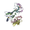


 Mass: 175.820 Da / Num. of mol.: 6 / Source method: obtained synthetically / Formula: Fe2S2
 Mass: 18.015 Da / Num. of mol.: 264 / Source method: isolated from a natural source / Formula: H2O
 Sample preparation
 / Beamline: BM14 / Wavelength: 0.98 Å
 Processing