 Movie
Movie Controller

[English] 日本語
 Yorodumi
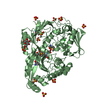
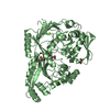
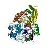
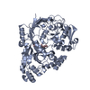
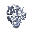
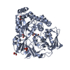
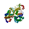
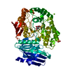
- PDB-4ofj: Crystal structure of NikA from Staphylococcus aureus in complex w... -
+ Open data

- Basic information
| Entry | Database: PDB / ID: 4ofj | ||||||
|---|---|---|---|---|---|---|---|
| Title | Crystal structure of NikA from Staphylococcus aureus in complex with Ni(L-His)2 | ||||||
 Components Components | Extracytoplasmic Nickel-Binding Protein SaNikA | ||||||
 Keywords Keywords | TRANSPORT PROTEIN / Extracytoplasmic / Nickel import / Metal transport / ABC-type importer / extracytoplasmic nickel-binding protein | ||||||
| Function / homology |  Function and homology information Function and homology informationnickel cation transport / peptide transport / peptide transmembrane transporter activity / ATP-binding cassette (ABC) transporter complex / transmembrane transport / periplasmic space Similarity search - Function | ||||||
| Biological species |   Staphylococcus aureus (bacteria) Staphylococcus aureus (bacteria) | ||||||
| Method |  X-RAY DIFFRACTION / SYNCHROTRON / MOLECULAR REPLACEMENT / Resolution: 1.7 Å X-RAY DIFFRACTION / SYNCHROTRON / MOLECULAR REPLACEMENT / Resolution: 1.7 Å | ||||||
 Authors Authors | Lebrette, H. / Cavazza, C. | ||||||
 Citation Citation | Journal: Metallomics / Year: 2015 Title: Novel insights into nickel import in Staphylococcus aureus: the positive role of free histidine and structural characterization of a new thiazolidine-type nickel chelator. Authors: Lebrette, H. / Borezee-Durant, E. / Martin, L. / Richaud, P. / Boeri Erba, E. / Cavazza, C. | ||||||
| History |
|
- Structure visualization
| Structure viewer | Molecule: MolmilJmol/JSmol |
|---|
- Downloads & links
-Download
| PDBx/mmCIF format | 4ofj.cif.gz | 212.4 KB | Display | PDBx/mmCIF format |
|---|---|---|---|---|
| PDB format | pdb4ofj.ent.gz | 169.4 KB | Display | PDB format |
| PDBx/mmJSON format | 4ofj.json.gz | Tree view | PDBx/mmJSON format | |
| Others |  Other downloads Other downloads |
-Validation report
| Arichive directory | https://data.pdbj.org/pub/pdb/validation_reports/of/4ofjftp://data.pdbj.org/pub/pdb/validation_reports/of/4ofj | HTTPS FTP |
|---|
-Related structure data
| Related structure data |  4xknC 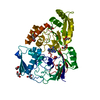 4xkpC 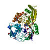 4xkqC 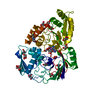 4xkrC 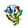 3rqtS S: Starting model for refinement C: citing same article ( |
|---|---|
| Similar structure data |


-Links
 PDBj
PDBj


- Assembly
| Deposited unit | 
| ||||||||
|---|---|---|---|---|---|---|---|---|---|
| 1 |
| ||||||||
| Unit cell |
|
-Components
-Protein , 1 types, 1 molecules A
| #1: Protein | Mass: 53545.285 Da / Num. of mol.: 1 Source method: isolated from a genetically manipulated source Source: (gene. exp.) Staphylococcus aureus (bacteria) / Strain: subsp. aureus COL / Gene: SACOL0217 / Production host: |
|---|
-Non-polymers , 5 types, 436 molecules 


| #2: Chemical | ChemComp-NI /  Mass: 58.693 Da / Num. of mol.: 1 / Source method: obtained synthetically / Formula: Ni Mass: 58.693 Da / Num. of mol.: 1 / Source method: obtained synthetically / Formula: Ni | ||||||
|---|---|---|---|---|---|---|---|
| #3: Chemical |  Type: L-peptide linking / Mass: 156.162 Da / Num. of mol.: 2 / Source method: obtained synthetically / Formula: C6H10N3O2 Type: L-peptide linking / Mass: 156.162 Da / Num. of mol.: 2 / Source method: obtained synthetically / Formula: C6H10N3O2#4: Chemical | ChemComp-EPE / |  Mass: 238.305 Da / Num. of mol.: 1 / Source method: obtained synthetically / Formula: C8H18N2O4S / Comment: pH buffer*YM Mass: 238.305 Da / Num. of mol.: 1 / Source method: obtained synthetically / Formula: C8H18N2O4S / Comment: pH buffer*YM#5: Chemical | ChemComp-GOL /  Mass: 92.094 Da / Num. of mol.: 17 / Source method: obtained synthetically / Formula: C3H8O3 Mass: 92.094 Da / Num. of mol.: 17 / Source method: obtained synthetically / Formula: C3H8O3#6: Water | ChemComp-HOH / | Mass: 18.015 Da / Num. of mol.: 415 / Source method: isolated from a natural source / Formula: H2O |
-Experimental details
-Experiment
| Experiment | Method: X-RAY DIFFRACTION / Number of used crystals: 1 |
|---|
- Sample preparation
| Crystal | Density Matthews: 2.26 Å3/Da / Density % sol: 45.66 % |
|---|---|
| Crystal grow | Temperature: 293 K / Method: vapor diffusion, hanging drop / pH: 7 Details: 27 % PEG 3350, 0.1 M HEPES pH 7.0, VAPOR DIFFUSION, HANGING DROP, temperature 293K |
-Data collection
| Diffraction source | Source: SYNCHROTRON / Site: ESRF  / Beamline: ID29 / Wavelength: 0.984 Å / Beamline: ID29 / Wavelength: 0.984 Å |
|---|---|
| Detector | Type: DECTRIS PILATUS 6M / Detector: PIXEL / Date: Nov 8, 2012 |
| Radiation | Protocol: SINGLE WAVELENGTH / Monochromatic (M) / Laue (L): M / Scattering type: x-ray |
| Radiation wavelength | Wavelength: 0.984 Å / Relative weight: 1 |
| Reflection | Resolution: 1.7→45.69 Å / Num. all: 54220 / Num. obs: 53990 / % possible obs: 99.6 % / Observed criterion σ(I): -3 / Redundancy: 7.2 % / Rsym value: 0.079 / Net I/σ(I): 17.04 |
| Reflection shell | Resolution: 1.7→1.8 Å / Redundancy: 7.4 % / Mean I/σ(I) obs: 4.23 / Num. unique all: 8418 / Rsym value: 0.652 / % possible all: 99.4 |
- Processing
| Software |
| ||||||||||||||||||||||||||||||||||||||||||||||||||||||||||||||||||||||||||||||||||||||||||||||||||||||||||||||||||||||||||||||||||||||||||||
|---|---|---|---|---|---|---|---|---|---|---|---|---|---|---|---|---|---|---|---|---|---|---|---|---|---|---|---|---|---|---|---|---|---|---|---|---|---|---|---|---|---|---|---|---|---|---|---|---|---|---|---|---|---|---|---|---|---|---|---|---|---|---|---|---|---|---|---|---|---|---|---|---|---|---|---|---|---|---|---|---|---|---|---|---|---|---|---|---|---|---|---|---|---|---|---|---|---|---|---|---|---|---|---|---|---|---|---|---|---|---|---|---|---|---|---|---|---|---|---|---|---|---|---|---|---|---|---|---|---|---|---|---|---|---|---|---|---|---|---|---|---|
| Refinement | Method to determine structure: MOLECULAR REPLACEMENT Starting model: PDB ENTRY 3RQT Resolution: 1.7→45.689 Å / SU ML: 0.21 / σ(F): 1.36 / Phase error: 16.57 / Stereochemistry target values: ML
| ||||||||||||||||||||||||||||||||||||||||||||||||||||||||||||||||||||||||||||||||||||||||||||||||||||||||||||||||||||||||||||||||||||||||||||
| Solvent computation | Shrinkage radii: 0.9 Å / VDW probe radii: 1.11 Å / Solvent model: FLAT BULK SOLVENT MODEL | ||||||||||||||||||||||||||||||||||||||||||||||||||||||||||||||||||||||||||||||||||||||||||||||||||||||||||||||||||||||||||||||||||||||||||||
| Refinement step | Cycle: LAST / Resolution: 1.7→45.689 Å
| ||||||||||||||||||||||||||||||||||||||||||||||||||||||||||||||||||||||||||||||||||||||||||||||||||||||||||||||||||||||||||||||||||||||||||||
| Refine LS restraints |
| ||||||||||||||||||||||||||||||||||||||||||||||||||||||||||||||||||||||||||||||||||||||||||||||||||||||||||||||||||||||||||||||||||||||||||||
| LS refinement shell |
| ||||||||||||||||||||||||||||||||||||||||||||||||||||||||||||||||||||||||||||||||||||||||||||||||||||||||||||||||||||||||||||||||||||||||||||
| Refinement TLS params. | Method: refined / Refine-ID: X-RAY DIFFRACTION
| ||||||||||||||||||||||||||||||||||||||||||||||||||||||||||||||||||||||||||||||||||||||||||||||||||||||||||||||||||||||||||||||||||||||||||||
| Refinement TLS group |
|
