 Movie
Movie Controller

[English] 日本語
 Yorodumi
- PDB-6tfx: Structure in P21 form of the PBP/SBP MoaA in complex with mannopi... -
+ Open data

- Basic information
| Entry | Database: PDB / ID: 6tfx | ||||||
|---|---|---|---|---|---|---|---|
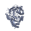
| Title | Structure in P21 form of the PBP/SBP MoaA in complex with mannopinic acid from A.tumefacien R10 | ||||||
 Components Components | ABC transporter substrate-binding protein | ||||||
 Keywords Keywords | TRANSPORT PROTEIN / Solute binding protein / periplasmic binding protein | ||||||
| Function / homology |  Function and homology information Function and homology informationpeptide transport / peptide transmembrane transporter activity / ATP-binding cassette (ABC) transporter complex / outer membrane-bounded periplasmic space / metal ion binding Similarity search - Function | ||||||
| Biological species |  Rhizobium radiobacter (Agrobacterium genomosp. 4) Rhizobium radiobacter (Agrobacterium genomosp. 4) | ||||||
| Method |  X-RAY DIFFRACTION / SYNCHROTRON / MOLECULAR REPLACEMENT / Resolution: 1.56 Å X-RAY DIFFRACTION / SYNCHROTRON / MOLECULAR REPLACEMENT / Resolution: 1.56 Å | ||||||
 Authors Authors | Morera, S. / Vigouroux, A. | ||||||
 Citation Citation | Journal: Biochem.J. / Year: 2020 Title: Import pathways of the mannityl-opines into the bacterial pathogen Agrobacterium tumefaciens: structural, affinity and in vivo approaches. Authors: Vigouroux, A. / Dore, J. / Marty, L. / Aumont-Nicaise, M. / Legrand, P. / Dessaux, Y. / Vial, L. / Morera, S. | ||||||
| History |
|
- Structure visualization
| Structure viewer | Molecule: MolmilJmol/JSmol |
|---|
- Downloads & links
-Download
| PDBx/mmCIF format | 6tfx.cif.gz | 406.4 KB | Display | PDBx/mmCIF format |
|---|---|---|---|---|
| PDB format | pdb6tfx.ent.gz | 328.4 KB | Display | PDB format |
| PDBx/mmJSON format | 6tfx.json.gz | Tree view | PDBx/mmJSON format | |
| Others |  Other downloads Other downloads |
-Validation report
| Arichive directory | https://data.pdbj.org/pub/pdb/validation_reports/tf/6tfxftp://data.pdbj.org/pub/pdb/validation_reports/tf/6tfx | HTTPS FTP |
|---|
-Related structure data
| Related structure data |  6tfqSC  6tfsC  6tg2C  6tg3C S: Starting model for refinement C: citing same article ( |
|---|---|
| Similar structure data |


-Links
 PDBj
PDBj- Assembly
| Deposited unit | 
| ||||||||
|---|---|---|---|---|---|---|---|---|---|
| 1 | 
| ||||||||
| 2 | 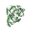
| ||||||||
| Unit cell |
|
-Components
-Protein , 1 types, 2 molecules AB
| #1: Protein | Mass: 55446.348 Da / Num. of mol.: 2 Source method: isolated from a genetically manipulated source Source: (gene. exp.) Rhizobium radiobacter (Agrobacterium genomosp. 4)Gene: moaA, ddpA, A6U90_18755, At1D1609_52430, AtA6_55190 / Production host: |
|---|
-Non-polymers , 5 types, 832 molecules 


| #2: Chemical |  Mass: 309.270 Da / Num. of mol.: 2 / Source method: obtained synthetically / Formula: C11H19NO9 / Feature type: SUBJECT OF INVESTIGATION Mass: 309.270 Da / Num. of mol.: 2 / Source method: obtained synthetically / Formula: C11H19NO9 / Feature type: SUBJECT OF INVESTIGATION#3: Chemical |  Mass: 62.068 Da / Num. of mol.: 3 / Source method: obtained synthetically / Formula: C2H6O2 Mass: 62.068 Da / Num. of mol.: 3 / Source method: obtained synthetically / Formula: C2H6O2#4: Chemical | ChemComp-PEG / |  Mass: 106.120 Da / Num. of mol.: 1 / Source method: obtained synthetically / Formula: C4H10O3 Mass: 106.120 Da / Num. of mol.: 1 / Source method: obtained synthetically / Formula: C4H10O3#5: Chemical | ChemComp-MG /  Mass: 24.305 Da / Num. of mol.: 4 / Source method: obtained synthetically / Formula: Mg Mass: 24.305 Da / Num. of mol.: 4 / Source method: obtained synthetically / Formula: Mg#6: Water | ChemComp-HOH / | Mass: 18.015 Da / Num. of mol.: 822 / Source method: isolated from a natural source / Formula: H2O |
|---|
-Details
| Has ligand of interest | Y |
|---|
-Experimental details
-Experiment
| Experiment | Method: X-RAY DIFFRACTION / Number of used crystals: 1 |
|---|
- Sample preparation
| Crystal | Density Matthews: 2.2 Å3/Da / Density % sol: 44.06 % |
|---|---|
| Crystal grow | Temperature: 292 K / Method: vapor diffusion, hanging drop / pH: 6.5 / Details: PEG 4K, 100 mM MES |
-Data collection
| Diffraction | Mean temperature: 100 K / Serial crystal experiment: N |
|---|---|
| Diffraction source | Source: SYNCHROTRON / Site: SOLEIL  / Beamline: PROXIMA 2 / Wavelength: 1 Å / Beamline: PROXIMA 2 / Wavelength: 1 Å |
| Detector | Type: DECTRIS EIGER2 X 9M / Detector: PIXEL / Date: Sep 6, 2018 |
| Radiation | Protocol: SINGLE WAVELENGTH / Monochromatic (M) / Laue (L): M / Scattering type: x-ray |
| Radiation wavelength | Wavelength: 1 Å / Relative weight: 1 |
| Reflection | Resolution: 1.56→47.56 Å / Num. obs: 134434 / % possible obs: 99.5 % / Redundancy: 6.7 % / CC1/2: 0.998 / Rmerge(I) obs: 0.094 / Rpim(I) all: 0.04 / Net I/σ(I): 9.4 |
| Reflection shell | Resolution: 1.56→1.66 Å / Rmerge(I) obs: 0.947 / Num. unique obs: 21895 / CC1/2: 0.646 |
- Processing
| Software |
| ||||||||||||||||||||||||||||||||||||||||||||||||||||||||||||||||||||||||||||||||||||||||||||||||||||||||||||
|---|---|---|---|---|---|---|---|---|---|---|---|---|---|---|---|---|---|---|---|---|---|---|---|---|---|---|---|---|---|---|---|---|---|---|---|---|---|---|---|---|---|---|---|---|---|---|---|---|---|---|---|---|---|---|---|---|---|---|---|---|---|---|---|---|---|---|---|---|---|---|---|---|---|---|---|---|---|---|---|---|---|---|---|---|---|---|---|---|---|---|---|---|---|---|---|---|---|---|---|---|---|---|---|---|---|---|---|---|---|
| Refinement | Method to determine structure: MOLECULAR REPLACEMENT Starting model: 6TFQ Resolution: 1.56→47.56 Å / Cor.coef. Fo:Fc: 0.968 / Cor.coef. Fo:Fc free: 0.96 / SU R Cruickshank DPI: 0.075 / Cross valid method: THROUGHOUT / σ(F): 0 / SU R Blow DPI: 0.076 / SU Rfree Blow DPI: 0.073 / SU Rfree Cruickshank DPI: 0.072
| ||||||||||||||||||||||||||||||||||||||||||||||||||||||||||||||||||||||||||||||||||||||||||||||||||||||||||||
| Displacement parameters | Biso max: 110.6 Å2 / Biso mean: 24.56 Å2 / Biso min: 10.68 Å2
| ||||||||||||||||||||||||||||||||||||||||||||||||||||||||||||||||||||||||||||||||||||||||||||||||||||||||||||
| Refine analyze | Luzzati coordinate error obs: 0.18 Å | ||||||||||||||||||||||||||||||||||||||||||||||||||||||||||||||||||||||||||||||||||||||||||||||||||||||||||||
| Refinement step | Cycle: final / Resolution: 1.56→47.56 Å
| ||||||||||||||||||||||||||||||||||||||||||||||||||||||||||||||||||||||||||||||||||||||||||||||||||||||||||||
| Refine LS restraints |
| ||||||||||||||||||||||||||||||||||||||||||||||||||||||||||||||||||||||||||||||||||||||||||||||||||||||||||||
| LS refinement shell | Resolution: 1.56→1.58 Å / Rfactor Rfree error: 0 / Total num. of bins used: 50
| ||||||||||||||||||||||||||||||||||||||||||||||||||||||||||||||||||||||||||||||||||||||||||||||||||||||||||||
| Refinement TLS params. | Method: refined / Refine-ID: X-RAY DIFFRACTION
| ||||||||||||||||||||||||||||||||||||||||||||||||||||||||||||||||||||||||||||||||||||||||||||||||||||||||||||
| Refinement TLS group |
|
