- PDB-3rqt: 1.5 Angstrom Crystal Structure of the Complex of Ligand Binding C... -
+
Open data
ID or keywords:
Loading...
-
Basic information
Entry
Database: PDB / ID: 3rqt
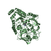
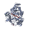
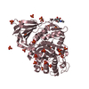
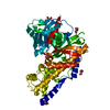
Title
1.5 Angstrom Crystal Structure of the Complex of Ligand Binding Component of ABC-type Import System from Staphylococcus aureus with Nickel and two Histidines
Components
Putative uncharacterized protein
Keywords
UNKNOWN FUNCTION / Ligand Binding Component / ABC-type Import System / Nickel / single or di-peptides / Structural Genomics / Center for Structural Genomics of Infectious Diseases / CSGID
Function / homology
Function and homology information
nickel cation transport / peptide transport / peptide transmembrane transporter activity / ATP-binding cassette (ABC) transporter complex / periplasmic space Similarity search - Function
: / Dipeptide-binding Protein; domain 3 / Dipeptide-binding Protein; Domain 3 / Peptide/nickel binding protein, MppA-type / Solute-binding protein family 5 domain / Solute-binding protein family 5 / Bacterial extracellular solute-binding proteins, family 5 Middle / Periplasmic binding protein-like II / D-Maltodextrin-Binding Protein; domain 2 / Prokaryotic membrane lipoprotein lipid attachment site profile. ...: / Dipeptide-binding Protein; domain 3 / Dipeptide-binding Protein; Domain 3 / Peptide/nickel binding protein, MppA-type / Solute-binding protein family 5 domain / Solute-binding protein family 5 / Bacterial extracellular solute-binding proteins, family 5 Middle / Periplasmic binding protein-like II / D-Maltodextrin-Binding Protein; domain 2 / Prokaryotic membrane lipoprotein lipid attachment site profile. / Roll / 3-Layer(aba) Sandwich / Alpha Beta Similarity search - Domain/homology
Type: MARMOSAIC 225 mm CCD / Detector: CCD / Date: Mar 17, 2011 / Details: Beryllium lenses
Radiation
Monochromator: Diamond / Protocol: SINGLE WAVELENGTH / Monochromatic (M) / Laue (L): M / Scattering type: x-ray
Radiation wavelength
Wavelength: 0.97872 Å / Relative weight: 1
Reflection
Resolution: 1.5→30 Å / Num. all: 77770 / Num. obs: 77770 / % possible obs: 99.9 % / Observed criterion σ(I): -3 / Redundancy: 4.9 % / Biso Wilson estimate: 12 Å2 / Rmerge(I) obs: 0.093 / Net I/σ(I): 21.5
Reflection shell
Resolution: 1.5→1.53 Å / Redundancy: 4.9 % / Rmerge(I) obs: 0.524 / Mean I/σ(I) obs: 3 / Num. unique all: 3817 / % possible all: 100
-
Processing
Software
Name
Version
Classification
Blu-Ice
Max
datacollection
HKL-3000
phasing
REFMAC
5.5.0102
refinement
HKL-3000
datareduction
HKL-3000
datascaling
Refinement
Method to determine structure: SAD / Resolution: 1.5→29.91 Å / Cor.coef. Fo:Fc: 0.972 / Cor.coef. Fo:Fc free: 0.961 / SU B: 2.124 / SU ML: 0.038 Isotropic thermal model: Thermal Factors Individually Refined Cross valid method: THROUGHOUT / ESU R Free: 0.067 / Stereochemistry target values: MAXIMUM LIKELIHOOD / Details: HYDROGENS HAVE BEEN ADDED IN THE RIDING POSITIONS
Rfactor
Num. reflection
% reflection
Selection details
Rfree
0.16971
3886
5 %
RANDOM
Rwork
0.14117
-
-
-
all
0.14259
73346
-
-
obs
0.14259
73346
99.83 %
-
Solvent computation
Ion probe radii: 0.8 Å / Shrinkage radii: 0.8 Å / VDW probe radii: 1.2 Å / Solvent model: BABINET MODEL WITH MASK
Displacement parameters
Biso mean: 15.24 Å2
Baniso -1
Baniso -2
Baniso -3
1-
0.17 Å2
-0 Å2
-0 Å2
2-
-
-0.11 Å2
0 Å2
3-
-
-
-0.06 Å2
Refinement step
Cycle: LAST / Resolution: 1.5→29.91 Å
Protein
Nucleic acid
Ligand
Solvent
Total
Num. atoms
3708
0
73
736
4517
Refine LS restraints
Refine-ID
Type
Dev ideal
Dev ideal target
Number
X-RAY DIFFRACTION
r_bond_refined_d
0.012
0.022
4272
X-RAY DIFFRACTION
r_bond_other_d
0.001
0.02
2953
X-RAY DIFFRACTION
r_angle_refined_deg
1.588
1.977
5813
X-RAY DIFFRACTION
r_angle_other_deg
0.944
3
7330
X-RAY DIFFRACTION
r_dihedral_angle_1_deg
3.938
5
548
X-RAY DIFFRACTION
r_dihedral_angle_2_deg
37.113
25.979
194
X-RAY DIFFRACTION
r_dihedral_angle_3_deg
9.079
15
832
X-RAY DIFFRACTION
r_dihedral_angle_4_deg
12.371
15
15
X-RAY DIFFRACTION
r_chiral_restr
0.107
0.2
626
X-RAY DIFFRACTION
r_gen_planes_refined
0.007
0.02
4818
X-RAY DIFFRACTION
r_gen_planes_other
0.002
0.02
767
X-RAY DIFFRACTION
r_mcbond_it
0.933
1.5
2605
X-RAY DIFFRACTION
r_mcbond_other
0.29
1.5
1026
X-RAY DIFFRACTION
r_mcangle_it
1.668
2
4288
X-RAY DIFFRACTION
r_scbond_it
2.766
3
1667
X-RAY DIFFRACTION
r_scangle_it
4.496
4.5
1525
LS refinement shell
Resolution: 1.5→1.539 Å / Total num. of bins used: 20
Rfactor
Num. reflection
% reflection
Rfree
0.206
299
-
Rwork
0.187
5302
-
obs
-
5302
99.63 %
Refinement TLS params.
Method: refined / Refine-ID: X-RAY DIFFRACTION
ID
L11 (°2)
L12 (°2)
L13 (°2)
L22 (°2)
L23 (°2)
L33 (°2)
S11 (Å °)
S12 (Å °)
S13 (Å °)
S21 (Å °)
S22 (Å °)
S23 (Å °)
S31 (Å °)
S32 (Å °)
S33 (Å °)
T11 (Å2)
T12 (Å2)
T13 (Å2)
T22 (Å2)
T23 (Å2)
T33 (Å2)
Origin x (Å)
Origin y (Å)
Origin z (Å)
1
0.3546
-0.1355
-0.2783
0.5864
0.3614
0.8852
-0.0304
0.0165
-0.0371
0.0464
-0.0171
-0.0009
0.0331
-0.0214
0.0475
0.0064
0.0026
0.0061
0.0165
0.0116
0.0189
42.1788
43.0308
13.8294
2
0.3238
-0.166
0.1107
1.0467
-0.0104
0.8396
-0.0413
-0.0218
0.0258
0.2234
-0.0098
-0.1489
-0.1043
0.164
0.0511
0.0612
-0.0201
-0.0418
0.0723
0.0046
0.043
50.2679
59.2932
29.8339
3
0.5641
0.0078
0.0727
0.9549
0.0499
0.7857
-0.0131
0.0432
0.0965
-0.0518
-0.0129
-0.0825
-0.1439
0.0753
0.026
0.0402
-0.0186
-0.0038
0.0128
0.013
0.0353
37.7524
76.8051
4.8922
4
0.4349
-0.0751
-0.0377
0.7339
0.1733
0.8769
0.0064
0.0193
0.0237
-0.0205
-0.0107
-0.0319
-0.0458
0.0181
0.0042
0.0029
-0.0009
-0.0007
0.0024
0.0032
0.0079
34.8704
66.7137
6.227
5
0.6459
0.536
0.2832
2.8093
0.3653
0.5816
-0.0117
0.103
-0.001
0.0515
0.0007
-0.3708
0.0076
0.2817
0.0109
0.0478
-0.0117
-0.0291
0.155
0.0253
0.1065
55.8941
56.0321
23.3273
Refinement TLS group
ID
Refine-ID
Refine TLS-ID
Auth asym-ID
Auth seq-ID
1
X-RAY DIFFRACTION
1
A
5 - 161
2
X-RAY DIFFRACTION
2
A
162 - 225
3
X-RAY DIFFRACTION
3
A
226 - 312
4
X-RAY DIFFRACTION
4
A
313 - 440
5
X-RAY DIFFRACTION
5
A
441 - 469
+
About Yorodumi
-
News
-
Feb 9, 2022. New format data for meta-information of EMDB entries
New format data for meta-information of EMDB entries
Version 3 of the EMDB header file is now the official format.
The previous official version 1.9 will be removed from the archive.
In the structure databanks used in Yorodumi, some data are registered as the other names, "COVID-19 virus" and "2019-nCoV". Here are the details of the virus and the list of structure data.
Jan 31, 2019. EMDB accession codes are about to change! (news from PDBe EMDB page)
EMDB accession codes are about to change! (news from PDBe EMDB page)
The allocation of 4 digits for EMDB accession codes will soon come to an end. Whilst these codes will remain in use, new EMDB accession codes will include an additional digit and will expand incrementally as the available range of codes is exhausted. The current 4-digit format prefixed with “EMD-” (i.e. EMD-XXXX) will advance to a 5-digit format (i.e. EMD-XXXXX), and so on. It is currently estimated that the 4-digit codes will be depleted around Spring 2019, at which point the 5-digit format will come into force.
The EM Navigator/Yorodumi systems omit the EMD- prefix.
Related info.:Q: What is EMD? / ID/Accession-code notation in Yorodumi/EM Navigator
Yorodumi is a browser for structure data from EMDB, PDB, SASBDB, etc.
This page is also the successor to EM Navigator detail page, and also detail information page/front-end page for Omokage search.
The word "yorodu" (or yorozu) is an old Japanese word meaning "ten thousand". "mi" (miru) is to see.
Related info.:EMDB / PDB / SASBDB / Comparison of 3 databanks / Yorodumi Search / Aug 31, 2016. New EM Navigator & Yorodumi / Yorodumi Papers / Jmol/JSmol / Function and homology information / Changes in new EM Navigator and Yorodumi
 Movie
Movie Controller

 Yorodumi
 Open data
 Basic information
 Components
 Keywords
 Function and homology information
 Staphylococcus aureus (bacteria)
 X-RAY DIFFRACTION /
  Authors
 Citation
 Structure visualization
 Downloads & links
 Other downloads
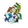


 PDBj
PDBj


 Assembly
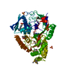


 Type: L-peptide linking / Mass: 156.162 Da / Num. of mol.: 2 / Source method: obtained synthetically / Formula: C6H10N3O2
 Mass: 58.693 Da / Num. of mol.: 1 / Source method: obtained synthetically / Formula: Ni
 Mass: 238.305 Da / Num. of mol.: 2 / Source method: obtained synthetically / Formula: C8H18N2O4S / Comment: pH buffer*YM
 Mass: 96.063 Da / Num. of mol.: 4 / Source method: obtained synthetically / Formula: SO4
 Sample preparation
 / Beamline: 21-ID-F / Wavelength: 0.97872 Å
 Processing