 Movie
Movie Controller

[English] 日本語
 Yorodumi
- PDB-4oec: Crystal structure of glycerophosphodiester phosphodiesterase from... -
+ Open data

- Basic information
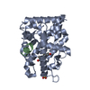
| Entry | Database: PDB / ID: 4oec | ||||||
|---|---|---|---|---|---|---|---|
| Title | Crystal structure of glycerophosphodiester phosphodiesterase from Thermococcus kodakarensis KOD1 | ||||||
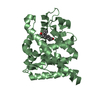
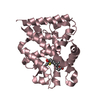
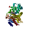
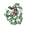
 Components Components | Glycerophosphoryl diester phosphodiesterase | ||||||
 Keywords Keywords | HYDROLASE / TIM Barrel | ||||||
| Function / homology |  Function and homology information Function and homology informationphosphoric diester hydrolase activity / lipid metabolic process / metal ion binding Similarity search - Function | ||||||
| Biological species |   Thermococcus kodakarensis (archaea) Thermococcus kodakarensis (archaea) | ||||||
| Method |  X-RAY DIFFRACTION / SYNCHROTRON / MOLECULAR REPLACEMENT / molecular replacement / Resolution: 1.9 Å X-RAY DIFFRACTION / SYNCHROTRON / MOLECULAR REPLACEMENT / molecular replacement / Resolution: 1.9 Å | ||||||
 Authors Authors | Atsuta, Y. / You, D.J. / Takano, K. / Koga, Y. / Kanaya, S. | ||||||
 Citation Citation | Journal: To be Published Title: Crystal structure of glycerophosphodiester phosphodiesterase from Thermococcus kodakarensis KOD1 Authors: Atsuta, Y. / You, D.J. / Takano, K. / Koga, Y. / Kanaya, S. | ||||||
| History |
|
- Structure visualization
| Structure viewer | Molecule: MolmilJmol/JSmol |
|---|
- Downloads & links
-Download
| PDBx/mmCIF format | 4oec.cif.gz | 220.4 KB | Display | PDBx/mmCIF format |
|---|---|---|---|---|
| PDB format | pdb4oec.ent.gz | 176.7 KB | Display | PDB format |
| PDBx/mmJSON format | 4oec.json.gz | Tree view | PDBx/mmJSON format | |
| Others |  Other downloads Other downloads |
-Validation report
| Summary document | 4oec_validation.pdf.gz | 450.8 KB | Display | wwPDB validaton report |
|---|---|---|---|---|
| Full document | 4oec_full_validation.pdf.gz | 463.2 KB | Display | |
| Data in XML | 4oec_validation.xml.gz | 43.6 KB | Display | |
| Data in CIF | 4oec_validation.cif.gz | 64 KB | Display | |
| Arichive directory | https://data.pdbj.org/pub/pdb/validation_reports/oe/4oecftp://data.pdbj.org/pub/pdb/validation_reports/oe/4oec | HTTPS FTP |
-Related structure data
| Related structure data |  2otdS S: Starting model for refinement |
|---|---|
| Similar structure data |

-Links
 PDBj
PDBj- Assembly
| Deposited unit | 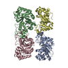
| ||||||||
|---|---|---|---|---|---|---|---|---|---|
| 1 | 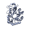
| ||||||||
| 2 | 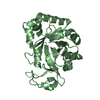
| ||||||||
| 3 | 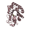
| ||||||||
| 4 | 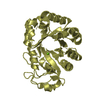
| ||||||||
| Unit cell |
|
-Components
| #1: Protein | Mass: 28555.945 Da / Num. of mol.: 4 Source method: isolated from a genetically manipulated source Source: (gene. exp.) Thermococcus kodakarensis (archaea) / Strain: KOD1 / Gene: TK1397 / Plasmid: pET25b / Production host:  References: UniProt: Q5JGZ3, glycerophosphodiester phosphodiesterase #2: Chemical | ChemComp-MG /   Mass: 24.305 Da / Num. of mol.: 4 / Source method: obtained synthetically / Formula: Mg Mass: 24.305 Da / Num. of mol.: 4 / Source method: obtained synthetically / Formula: Mg#3: Water | ChemComp-HOH / |  Mass: 18.015 Da / Num. of mol.: 693 / Source method: isolated from a natural source / Formula: H2O Mass: 18.015 Da / Num. of mol.: 693 / Source method: isolated from a natural source / Formula: H2O |
|---|
-Experimental details
-Experiment
| Experiment | Method: X-RAY DIFFRACTION / Number of used crystals: 1 |
|---|
- Sample preparation
| Crystal | Density Matthews: 2.17 Å3/Da / Density % sol: 43.28 % / Mosaicity: 0.507 ° / Mosaicity esd: 0.002 ° |
|---|---|
| Crystal grow | Temperature: 293 K / Method: vapor diffusion, sitting drop / pH: 8.5 Details: 30%(w/v) PEG4000, 0.2M Magnesium chloride hexahydrate, 0.1M Tris-HCl, pH 8.5, VAPOR DIFFUSION, SITTING DROP, temperature 293K |
-Data collection
| Diffraction | Mean temperature: 100 K | ||||||||||||||||||||||||||||||||||||||||||||||||||||||||||||||||||||||||||||||||||||||||||||||||||||||||||||||||||||||||||||||
|---|---|---|---|---|---|---|---|---|---|---|---|---|---|---|---|---|---|---|---|---|---|---|---|---|---|---|---|---|---|---|---|---|---|---|---|---|---|---|---|---|---|---|---|---|---|---|---|---|---|---|---|---|---|---|---|---|---|---|---|---|---|---|---|---|---|---|---|---|---|---|---|---|---|---|---|---|---|---|---|---|---|---|---|---|---|---|---|---|---|---|---|---|---|---|---|---|---|---|---|---|---|---|---|---|---|---|---|---|---|---|---|---|---|---|---|---|---|---|---|---|---|---|---|---|---|---|---|
| Diffraction source | Source: SYNCHROTRON / Site: SPring-8  / Beamline: BL44XU / Wavelength: 0.9 Å / Beamline: BL44XU / Wavelength: 0.9 Å | ||||||||||||||||||||||||||||||||||||||||||||||||||||||||||||||||||||||||||||||||||||||||||||||||||||||||||||||||||||||||||||||
| Detector | Type: BRUKER SMART 6500 / Detector: CCD / Date: Jul 4, 2012 | ||||||||||||||||||||||||||||||||||||||||||||||||||||||||||||||||||||||||||||||||||||||||||||||||||||||||||||||||||||||||||||||
| Radiation | Monochromator: horizontal focusing mirror / Protocol: SINGLE WAVELENGTH / Monochromatic (M) / Laue (L): M / Scattering type: x-ray | ||||||||||||||||||||||||||||||||||||||||||||||||||||||||||||||||||||||||||||||||||||||||||||||||||||||||||||||||||||||||||||||
| Radiation wavelength | Wavelength: 0.9 Å / Relative weight: 1 | ||||||||||||||||||||||||||||||||||||||||||||||||||||||||||||||||||||||||||||||||||||||||||||||||||||||||||||||||||||||||||||||
| Reflection | Resolution: 1.9→50 Å / Num. obs: 79998 / % possible obs: 99.8 % / Observed criterion σ(F): 0 / Observed criterion σ(I): -3 / Redundancy: 13.6 % / Rmerge(I) obs: 0.142 / Rsym value: 0.142 / Net I/σ(I): 23.493 | ||||||||||||||||||||||||||||||||||||||||||||||||||||||||||||||||||||||||||||||||||||||||||||||||||||||||||||||||||||||||||||||
| Reflection shell |
|
-Phasing
| Phasing | Method: molecular replacement |
|---|
- Processing
| Software |
| |||||||||||||||||||||||||||||||||||||||||||||||||||||||||||||||||
|---|---|---|---|---|---|---|---|---|---|---|---|---|---|---|---|---|---|---|---|---|---|---|---|---|---|---|---|---|---|---|---|---|---|---|---|---|---|---|---|---|---|---|---|---|---|---|---|---|---|---|---|---|---|---|---|---|---|---|---|---|---|---|---|---|---|---|
| Refinement | Method to determine structure: MOLECULAR REPLACEMENT Starting model: 2otd Resolution: 1.9→50 Å / Cor.coef. Fo:Fc: 0.955 / Cor.coef. Fo:Fc free: 0.93 / WRfactor Rfree: 0.2426 / WRfactor Rwork: 0.1901 / FOM work R set: 0.8283 / SU B: 3.673 / SU ML: 0.111 / SU R Cruickshank DPI: 0.1734 / SU Rfree: 0.1639 / Cross valid method: THROUGHOUT / σ(F): 0 / ESU R: 0.173 / ESU R Free: 0.164 / Stereochemistry target values: MAXIMUM LIKELIHOOD Details: HYDROGENS HAVE BEEN ADDED IN THE RIDING POSITIONS U VALUES: REFINED INDIVIDUALLY
| |||||||||||||||||||||||||||||||||||||||||||||||||||||||||||||||||
| Solvent computation | Ion probe radii: 0.8 Å / Shrinkage radii: 0.8 Å / VDW probe radii: 1.4 Å / Solvent model: MASK | |||||||||||||||||||||||||||||||||||||||||||||||||||||||||||||||||
| Displacement parameters | Biso max: 70.6 Å2 / Biso mean: 30.063 Å2 / Biso min: 13.6 Å2
| |||||||||||||||||||||||||||||||||||||||||||||||||||||||||||||||||
| Refinement step | Cycle: LAST / Resolution: 1.9→50 Å
| |||||||||||||||||||||||||||||||||||||||||||||||||||||||||||||||||
| Refine LS restraints |
| |||||||||||||||||||||||||||||||||||||||||||||||||||||||||||||||||
| LS refinement shell | Resolution: 1.9→1.943 Å / Total num. of bins used: 20
|
