 Movie
Movie Controller

[English] 日本語
 Yorodumi
- PDB-4o29: PROTEIN-L-ISOASPARTATE O-METHYLTRANSFERASE from Pyrobaculum aerop... -
+ Open data

- Basic information
| Entry | Database: PDB / ID: 4o29 | ||||||
|---|---|---|---|---|---|---|---|
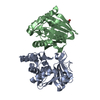
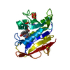
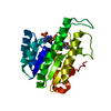
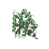
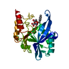
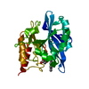
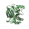
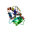
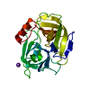
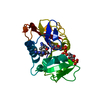
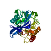
| Title | PROTEIN-L-ISOASPARTATE O-METHYLTRANSFERASE from Pyrobaculum aerophilum in COMPLEX WITH S-ADENOSYL-L-HOMOCYSTEINE | ||||||
 Components Components | Protein-L-isoaspartate O-methyltransferase | ||||||
 Keywords Keywords | TRANSFERASE / ROSSMANN METHYLTRANSFERASE / PROTEIN REPAIR ISOMERIZATION | ||||||
| Function / homology |  Function and homology information Function and homology informationprotein-L-isoaspartate(D-aspartate) O-methyltransferase / protein-L-isoaspartate (D-aspartate) O-methyltransferase activity / protein repair / protein modification process / methylation / cytoplasm Similarity search - Function | ||||||
| Biological species |   Pyrobaculum aerophilum (archaea) Pyrobaculum aerophilum (archaea) | ||||||
| Method |  X-RAY DIFFRACTION / SYNCHROTRON / MOLECULAR REPLACEMENT / molecular replacement / Resolution: 2.9 Å X-RAY DIFFRACTION / SYNCHROTRON / MOLECULAR REPLACEMENT / molecular replacement / Resolution: 2.9 Å | ||||||
 Authors Authors | Sawaya, M.R. / Yeates, T.O. / Griffith, S.C. | ||||||
 Citation Citation | Journal: Thesis / Year: 2002 Title: Structure of L-isoaspartyl (D-aspartyl) Methyltransferase Authors: Griffith, S.C. #1: Journal: J.Mol.Biol. / Year: 2001Title: Crystal structure of a protein repair methyltransferase from pyrococcus furiosus with its l-isoaspartyl peptide substrate Authors: Griffith, S.C. / Sawaya, M.R. / Boutz, D.R. / Thapar, N. / Katz, J.E. / Clarke, S. / Yeates, T.O. | ||||||
| History |
|
- Structure visualization
| Structure viewer | Molecule: MolmilJmol/JSmol |
|---|
- Downloads & links
-Download
| PDBx/mmCIF format | 4o29.cif.gz | 97.4 KB | Display | PDBx/mmCIF format |
|---|---|---|---|---|
| PDB format | pdb4o29.ent.gz | 73.3 KB | Display | PDB format |
| PDBx/mmJSON format | 4o29.json.gz | Tree view | PDBx/mmJSON format | |
| Others |  Other downloads Other downloads |
-Validation report
| Summary document | 4o29_validation.pdf.gz | 667.5 KB | Display | wwPDB validaton report |
|---|---|---|---|---|
| Full document | 4o29_full_validation.pdf.gz | 669.3 KB | Display | |
| Data in XML | 4o29_validation.xml.gz | 9.7 KB | Display | |
| Data in CIF | 4o29_validation.cif.gz | 12.1 KB | Display | |
| Arichive directory | https://data.pdbj.org/pub/pdb/validation_reports/o2/4o29ftp://data.pdbj.org/pub/pdb/validation_reports/o2/4o29 | HTTPS FTP |
-Related structure data
| Related structure data |  1jg1S S: Starting model for refinement |
|---|---|
| Similar structure data |
-Links
 PDBj
PDBj- Assembly
| Deposited unit | 
| ||||||||
|---|---|---|---|---|---|---|---|---|---|
| 1 |
| ||||||||
| Unit cell |
|
-Components
| #1: Protein | Mass: 23236.578 Da / Num. of mol.: 1 Source method: isolated from a genetically manipulated source Source: (gene. exp.) Pyrobaculum aerophilum (archaea) / Strain: IM2 / Gene: PAE0701, pcm, PIMT / Plasmid: pTrcHis2-TOPO / Production host:  References: UniProt: Q8ZYN0, protein-L-isoaspartate(D-aspartate) O-methyltransferase |
|---|---|
| #2: Chemical | ChemComp-SAH /   Type: L-peptide linking / Mass: 384.411 Da / Num. of mol.: 1 / Source method: obtained synthetically / Formula: C14H20N6O5S Type: L-peptide linking / Mass: 384.411 Da / Num. of mol.: 1 / Source method: obtained synthetically / Formula: C14H20N6O5S |
| #3: Water | ChemComp-HOH /  Mass: 18.015 Da / Num. of mol.: 2 / Source method: isolated from a natural source / Formula: H2O Mass: 18.015 Da / Num. of mol.: 2 / Source method: isolated from a natural source / Formula: H2O |
-Experimental details
-Experiment
| Experiment | Method: X-RAY DIFFRACTION / Number of used crystals: 1 |
|---|
- Sample preparation
| Crystal | Density Matthews: 1.85 Å3/Da / Density % sol: 33.34 % |
|---|---|
| Crystal grow | Temperature: 298 K / Method: vapor diffusion, hanging drop / pH: 7.5 Details: 70% MPD, 0.1M HEPES pH 7.5, vapor diffusion, hanging drop, temperature 298K |
-Data collection
| Diffraction | Mean temperature: 100 K | |||||||||||||||||||||||||||||||||||||||||||||||||||||||||||||||||||||||||||||||||||||||||||||||||||||||||||||||||||||||||||||||||||||||||||||||||||
|---|---|---|---|---|---|---|---|---|---|---|---|---|---|---|---|---|---|---|---|---|---|---|---|---|---|---|---|---|---|---|---|---|---|---|---|---|---|---|---|---|---|---|---|---|---|---|---|---|---|---|---|---|---|---|---|---|---|---|---|---|---|---|---|---|---|---|---|---|---|---|---|---|---|---|---|---|---|---|---|---|---|---|---|---|---|---|---|---|---|---|---|---|---|---|---|---|---|---|---|---|---|---|---|---|---|---|---|---|---|---|---|---|---|---|---|---|---|---|---|---|---|---|---|---|---|---|---|---|---|---|---|---|---|---|---|---|---|---|---|---|---|---|---|---|---|---|---|---|
| Diffraction source | Source: SYNCHROTRON / Site: NSLS  / Beamline: X8C / Wavelength: 0.9789 Å / Beamline: X8C / Wavelength: 0.9789 Å | |||||||||||||||||||||||||||||||||||||||||||||||||||||||||||||||||||||||||||||||||||||||||||||||||||||||||||||||||||||||||||||||||||||||||||||||||||
| Detector | Type: ADSC QUANTUM 4 / Detector: CCD / Date: Apr 16, 1999 | |||||||||||||||||||||||||||||||||||||||||||||||||||||||||||||||||||||||||||||||||||||||||||||||||||||||||||||||||||||||||||||||||||||||||||||||||||
| Radiation | Monochromator: MIRROR / Protocol: SINGLE WAVELENGTH / Monochromatic (M) / Laue (L): M / Scattering type: x-ray | |||||||||||||||||||||||||||||||||||||||||||||||||||||||||||||||||||||||||||||||||||||||||||||||||||||||||||||||||||||||||||||||||||||||||||||||||||
| Radiation wavelength | Wavelength: 0.9789 Å / Relative weight: 1 | |||||||||||||||||||||||||||||||||||||||||||||||||||||||||||||||||||||||||||||||||||||||||||||||||||||||||||||||||||||||||||||||||||||||||||||||||||
| Reflection | Resolution: 2.9→65.22 Å / Num. all: 3881 / Num. obs: 3881 / % possible obs: 98.9 % / Observed criterion σ(I): -3 / Redundancy: 6.1 % / Biso Wilson estimate: 78.84 Å2 / Rmerge(I) obs: 0.171 / Net I/σ(I): 7.99 | |||||||||||||||||||||||||||||||||||||||||||||||||||||||||||||||||||||||||||||||||||||||||||||||||||||||||||||||||||||||||||||||||||||||||||||||||||
| Reflection shell | Diffraction-ID: 1
|
-Phasing
| Phasing | Method: molecular replacement | ||||||
|---|---|---|---|---|---|---|---|
| Phasing MR | Rfactor: 0.509 / Cor.coef. Fo:Fc: 0.41
|
- Processing
| Software |
| ||||||||||||||||||||||||||||||||||||||||
|---|---|---|---|---|---|---|---|---|---|---|---|---|---|---|---|---|---|---|---|---|---|---|---|---|---|---|---|---|---|---|---|---|---|---|---|---|---|---|---|---|---|
| Refinement | Method to determine structure: MOLECULAR REPLACEMENT Starting model: PDB ENTRY 1JG1 Resolution: 2.9→65.22 Å / Cor.coef. Fo:Fc: 0.9056 / Cor.coef. Fo:Fc free: 0.8308 / Occupancy max: 1 / Occupancy min: 1 / Cross valid method: THROUGHOUT / σ(F): 0 / Stereochemistry target values: Engh & Huber
| ||||||||||||||||||||||||||||||||||||||||
| Solvent computation | Bsol: 104.889 Å2 | ||||||||||||||||||||||||||||||||||||||||
| Displacement parameters | Biso max: 136.16 Å2 / Biso mean: 64.6522 Å2 / Biso min: 35 Å2
| ||||||||||||||||||||||||||||||||||||||||
| Refine analyze | Luzzati coordinate error obs: 0.443 Å | ||||||||||||||||||||||||||||||||||||||||
| Refinement step | Cycle: LAST / Resolution: 2.9→65.22 Å
| ||||||||||||||||||||||||||||||||||||||||
| Refine LS restraints |
| ||||||||||||||||||||||||||||||||||||||||
| LS refinement shell | Resolution: 2.9→3.24 Å / Total num. of bins used: 5
| ||||||||||||||||||||||||||||||||||||||||
| Refinement TLS params. | Method: refined / Origin x: 37.9205 Å / Origin y: 48.3813 Å / Origin z: 50.0303 Å
| ||||||||||||||||||||||||||||||||||||||||
| Refinement TLS group | Selection details: { A|* } | ||||||||||||||||||||||||||||||||||||||||
| Xplor file |
|
