 Movie
Movie Controller

[English] 日本語
 Yorodumi
- PDB-4nzt: Crystal structure of the antibody-binding region of Protein M (Pr... -
+ Open data

- Basic information
| Entry | Database: PDB / ID: 4nzt | ||||||
|---|---|---|---|---|---|---|---|
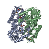
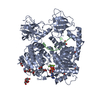
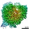
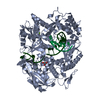
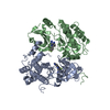
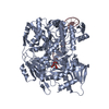
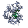
| Title | Crystal structure of the antibody-binding region of Protein M (Protein M TD) in complex with anti-infleunza hemagglutinin antibody CR9114 Fab | ||||||
 Components Components |
| ||||||
 Keywords Keywords | PROTEIN BINDING/IMMUNE SYSTEM / Leucine-rich repeat / broad antibody-binding / block antibody-antigen union / variable region / PROTEIN BINDING-IMMUNE SYSTEM complex | ||||||
| Function / homology |  Function and homology information Function and homology information | ||||||
| Biological species |  Mycoplasma genitalium (bacteria) Mycoplasma genitalium (bacteria) Homo sapiens (human) Homo sapiens (human) | ||||||
| Method |  X-RAY DIFFRACTION / SYNCHROTRON / MOLECULAR REPLACEMENT / Resolution: 2.497 Å X-RAY DIFFRACTION / SYNCHROTRON / MOLECULAR REPLACEMENT / Resolution: 2.497 Å | ||||||
 Authors Authors | Zhu, X. / Wilson, I.A. | ||||||
 Citation Citation | Journal: Science / Year: 2014 Title: A structurally distinct human mycoplasma protein that generically blocks antigen-antibody union. Authors: Rajesh K Grover / Xueyong Zhu / Travis Nieusma / Teresa Jones / Isabel Boreo / Amanda S MacLeod / Adam Mark / Sherry Niessen / Helen J Kim / Leopold Kong / Nacyra Assad-Garcia / Keehwan Kwon ...Authors: Rajesh K Grover / Xueyong Zhu / Travis Nieusma / Teresa Jones / Isabel Boreo / Amanda S MacLeod / Adam Mark / Sherry Niessen / Helen J Kim / Leopold Kong / Nacyra Assad-Garcia / Keehwan Kwon / Marta Chesi / Vaughn V Smider / Daniel R Salomon / Diane F Jelinek / Robert A Kyle / Richard B Pyles / John I Glass / Andrew B Ward / Ian A Wilson / Richard A Lerner /  Abstract: We report the discovery of a broadly reactive antibody-binding protein (Protein M) from human mycoplasma. The crystal structure of the ectodomain of transmembrane Protein M differs from other known ...We report the discovery of a broadly reactive antibody-binding protein (Protein M) from human mycoplasma. The crystal structure of the ectodomain of transmembrane Protein M differs from other known protein structures, as does its mechanism of antibody binding. Protein M binds with high affinity to all types of human and nonhuman immunoglobulin G, predominantly through attachment to the conserved portions of the variable region of the κ and λ light chains. Protein M blocks antibody-antigen union, likely because of its large C-terminal domain extending over the antibody-combining site, blocking entry to large antigens. Similar to the other immunoglobulin-binding proteins such as Protein A, Protein M as well as its orthologs in other Mycoplasma species could become invaluable reagents in the antibody field. | ||||||
| History |
|
- Structure visualization
| Structure viewer | Molecule: MolmilJmol/JSmol |
|---|
- Downloads & links
-Download
| PDBx/mmCIF format | 4nzt.cif.gz | 175.2 KB | Display | PDBx/mmCIF format |
|---|---|---|---|---|
| PDB format | pdb4nzt.ent.gz | 134.3 KB | Display | PDB format |
| PDBx/mmJSON format | 4nzt.json.gz | Tree view | PDBx/mmJSON format | |
| Others |  Other downloads Other downloads |
-Validation report
| Arichive directory | https://data.pdbj.org/pub/pdb/validation_reports/nz/4nztftp://data.pdbj.org/pub/pdb/validation_reports/nz/4nzt | HTTPS FTP |
|---|
-Related structure data
| Related structure data |  5834C  5835C  5836C  4nzrSC  4nzuC  4fqhS S: Starting model for refinement C: citing same article ( |
|---|---|
| Similar structure data |


-Links
 PDBj
PDBj

- Assembly
| Deposited unit | 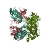
| ||||||||
|---|---|---|---|---|---|---|---|---|---|
| 1 |
| ||||||||
| Unit cell |
|
-Components
| #1: Protein | Mass: 46972.684 Da / Num. of mol.: 1 / Fragment: antibody-binding region (UNP residues 74-468) Source method: isolated from a genetically manipulated source Source: (gene. exp.) Mycoplasma genitalium (bacteria) / Strain: ATCC 33530 / G-37 / NCTC 10195 / Gene: MG281 / Production host: |
|---|---|
| #2: Antibody | Mass: 24377.166 Da / Num. of mol.: 1 / Fragment: Fab Source method: isolated from a genetically manipulated source Source: (gene. exp.) Homo sapiens (human) / Production host:  Trichoplusia ni (cabbage looper) Trichoplusia ni (cabbage looper) |
| #3: Antibody | Mass: 22840.172 Da / Num. of mol.: 1 / Fragment: Fab Source method: isolated from a genetically manipulated source Source: (gene. exp.) Homo sapiens (human) / Production host: Trichoplusia ni (cabbage looper) |
| #4: Water | ChemComp-HOH /  Mass: 18.015 Da / Num. of mol.: 270 / Source method: isolated from a natural source / Formula: H2O Mass: 18.015 Da / Num. of mol.: 270 / Source method: isolated from a natural source / Formula: H2O |
| Has protein modification | Y |
-Experimental details
-Experiment
| Experiment | Method: X-RAY DIFFRACTION / Number of used crystals: 1 |
|---|
- Sample preparation
| Crystal | Density Matthews: 2.15 Å3/Da / Density % sol: 42.68 % |
|---|---|
| Crystal grow | Temperature: 295 K / Method: vapor diffusion, sitting drop / pH: 6.29 Details: 0.1 M MES, pH 6.26, 6% MPD, VAPOR DIFFUSION, SITTING DROP, temperature 295K |
-Data collection
| Diffraction | Mean temperature: 100 K |
|---|---|
| Diffraction source | Source: SYNCHROTRON / Site: SSRL / Beamline: BL11-1 / Wavelength: 0.97945 Å |
| Detector | Type: DECTRIS PILATUS 6M / Detector: PIXEL / Date: Jul 18, 2013 |
| Radiation | Monochromator: Side scattering bent cube-root I-beam single crystal, asymmetric cut 4.965 degrees Protocol: SINGLE WAVELENGTH / Monochromatic (M) / Laue (L): M / Scattering type: x-ray |
| Radiation wavelength | Wavelength: 0.97945 Å / Relative weight: 1 |
| Reflection | Resolution: 2.497→50 Å / Num. obs: 27816 / % possible obs: 98 % / Observed criterion σ(I): -3 / Redundancy: 5 % / Rsym value: 0.13 / Net I/σ(I): 15.3 |
| Reflection shell | Resolution: 2.497→2.54 Å / Redundancy: 2.8 % / Mean I/σ(I) obs: 1.5 / Rsym value: 0.44 / % possible all: 84.5 |
- Processing
| Software |
| |||||||||||||||||||||||||||||||||||||||||||||||||||||||||||||||||||||||||||||
|---|---|---|---|---|---|---|---|---|---|---|---|---|---|---|---|---|---|---|---|---|---|---|---|---|---|---|---|---|---|---|---|---|---|---|---|---|---|---|---|---|---|---|---|---|---|---|---|---|---|---|---|---|---|---|---|---|---|---|---|---|---|---|---|---|---|---|---|---|---|---|---|---|---|---|---|---|---|---|
| Refinement | Method to determine structure: MOLECULAR REPLACEMENT Starting model: PDB ENTRIES 4NZR AND 4FQH Resolution: 2.497→41.727 Å / SU ML: 0.3 / Cross valid method: THROUGHOUT / σ(F): 1.36 / Phase error: 26.14 / Stereochemistry target values: ML
| |||||||||||||||||||||||||||||||||||||||||||||||||||||||||||||||||||||||||||||
| Solvent computation | Shrinkage radii: 0.9 Å / VDW probe radii: 1.11 Å / Solvent model: FLAT BULK SOLVENT MODEL | |||||||||||||||||||||||||||||||||||||||||||||||||||||||||||||||||||||||||||||
| Refinement step | Cycle: LAST / Resolution: 2.497→41.727 Å
| |||||||||||||||||||||||||||||||||||||||||||||||||||||||||||||||||||||||||||||
| Refine LS restraints |
| |||||||||||||||||||||||||||||||||||||||||||||||||||||||||||||||||||||||||||||
| LS refinement shell |
|
