- PDB-4nzj: Crystal structure of a putative alpha-galactosidase (BF1418) from... -
+
Open data
ID or keywords:
Loading...
-
Basic information
Entry
Database: PDB / ID: 4nzj
Title
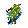
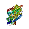
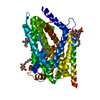
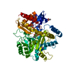
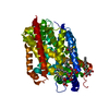
Crystal structure of a putative alpha-galactosidase (BF1418) from Bacteroides fragilis NCTC 9343 at 1.57 A resolution
Components
Putative alpha-galactosidase
Keywords
HYDROLASE / The catalytic TIM beta/alpha barrel domain accompanied by N-terminal Ig-like domain and C-terminal beta-sheet domain / Structural Genomics / Joint Center for Structural Genomics / JCSG / Protein Structure Initiative / PSI-BIOLOGY
Function / homology
Function and homology information
alpha-galactosidase / alpha-galactosidase activity / carbohydrate metabolic process / calcium ion binding / extracellular region / membrane Similarity search - Function
Alpha galactosidase, C-terminal beta sandwich domain / Alpha galactosidase C-terminal beta sandwich domain / Putative Ig domain / Alpha galactosidase A / Glycoside hydrolase, family 27 / Cadherin-like superfamily / Golgi alpha-mannosidase II / Glycosyl hydrolase, all-beta / Aldolase class I / Aldolase-type TIM barrel ...Alpha galactosidase, C-terminal beta sandwich domain / Alpha galactosidase C-terminal beta sandwich domain / Putative Ig domain / Alpha galactosidase A / Glycoside hydrolase, family 27 / Cadherin-like superfamily / Golgi alpha-mannosidase II / Glycosyl hydrolase, all-beta / Aldolase class I / Aldolase-type TIM barrel / Glycoside hydrolase superfamily / TIM Barrel / Alpha-Beta Barrel / Immunoglobulin-like fold / Immunoglobulins / Immunoglobulin-like / Sandwich / Mainly Beta / Alpha Beta Similarity search - Domain/homology
Mass: 18.015 Da / Num. of mol.: 480 / Source method: isolated from a natural source / Formula: H2O
Has protein modification
Y
Sequence details
THIS CONSTRUCT WAS EXPRESSED WITH AN N-TERMINAL PURIFICATION TAG MGSDKIHHHHHHENLYFQG. THE TAG WAS ...THIS CONSTRUCT WAS EXPRESSED WITH AN N-TERMINAL PURIFICATION TAG MGSDKIHHHHHHENLYFQG. THE TAG WAS REMOVED WITH TEV PROTEASE WITH TEV PROTEASE LEAVING ONLY A GLYCINE (0) FOLLOWED BY RESIDUES 25-500 OF THE TARGET SEQUENCE.
-
Experimental details
-
Experiment
Experiment
Method: X-RAY DIFFRACTION / Number of used crystals: 1
-
Sample preparation
Crystal
Density Matthews: 2.35 Å3/Da / Density % sol: 47.62 %
Resolution: 1.57→29.386 Å / Num. obs: 71815 / % possible obs: 99.1 % / Observed criterion σ(I): -3 / Biso Wilson estimate: 22.74 Å2 / Rmerge(I) obs: 0.076 / Net I/σ(I): 9.28
Reflection shell
Diffraction-ID: 1
Resolution (Å)
Highest resolution (Å)
Rmerge(I) obs
Mean I/σ(I) obs
Num. measured obs
Num. unique obs
% possible all
1.57-1.63
0.947
1.5
51478
13878
94.7
1.63-1.69
0.741
2.1
47845
12582
99.6
1.69-1.77
0.562
2.8
54384
14266
99.8
1.77-1.86
0.387
4
50606
13253
99.8
1.86-1.98
0.258
5.8
54180
14155
99.8
1.98-2.13
0.168
8.3
51543
13445
99.8
2.13-2.34
0.115
11.2
51874
13560
99.8
2.34-2.68
0.085
14.2
52781
13843
99.7
2.68-3.38
0.06
18.6
51503
13810
99.4
3.38
0.044
23.9
50734
13734
99.3
-
Phasing
Phasing
Method: MAD
-
Processing
Software
Name
Version
Classification
NB
MolProbity
3beta29
modelbuilding
PDB_EXTRACT
3.1
dataextraction
SHELX
phasing
SHARP
phasing
XSCALE
datascaling
REFMAC
5.7.0032
refinement
XDS
datareduction
SHELXD
phasing
Refinement
Method to determine structure: MAD / Resolution: 1.57→29.386 Å / Cor.coef. Fo:Fc: 0.975 / Cor.coef. Fo:Fc free: 0.973 / Occupancy max: 1 / Occupancy min: 0.1 / SU B: 2.612 / SU ML: 0.046 / Cross valid method: THROUGHOUT / σ(F): 0 / ESU R: 0.072 / ESU R Free: 0.068 Stereochemistry target values: MAXIMUM LIKELIHOOD WITH PHASES Details: 1. HYDROGENS HAVE BEEN ADDED IN THE RIDING POSITIONS. 2. ATOM RECORDS CONTAIN SUM OF TLS AND RESIDUAL B FACTORS. 3. ANISOU RECORDS CONTAIN SUM OF TLS AND RESIDUAL U FACTORS. 4. WATERS WERE ...Details: 1. HYDROGENS HAVE BEEN ADDED IN THE RIDING POSITIONS. 2. ATOM RECORDS CONTAIN SUM OF TLS AND RESIDUAL B FACTORS. 3. ANISOU RECORDS CONTAIN SUM OF TLS AND RESIDUAL U FACTORS. 4. WATERS WERE EXCLUDED FROM AUTOMATIC TLS ASSIGNMENT. 5. A MET-INHIBITION PROTOCOL WAS USED FOR SELENOMETHIONINE INCORPORATION DURING PROTEIN EXPRESSION. THE OCCUPANCY OF THE SE ATOMS IN THE MSE RESIDUES WAS REDUCED TO 0.75 FOR THE REDUCED SCATTERING POWER DUE TO PARTIAL S-MET INCORPORATION. 6. GLYCEROL (GOL) FROM THE CRYSTALLIZATION SOLUTION IS MODELED.
Rfactor
Num. reflection
% reflection
Selection details
Rfree
0.1685
3628
5.1 %
RANDOM
Rwork
0.1541
-
-
-
obs
0.1549
71742
99.75 %
-
Solvent computation
Ion probe radii: 0.8 Å / Shrinkage radii: 0.8 Å / VDW probe radii: 1.2 Å / Solvent model: BABINET MODEL WITH MASK
In the structure databanks used in Yorodumi, some data are registered as the other names, "COVID-19 virus" and "2019-nCoV". Here are the details of the virus and the list of structure data.
Jan 31, 2019. EMDB accession codes are about to change! (news from PDBe EMDB page)
EMDB accession codes are about to change! (news from PDBe EMDB page)
The allocation of 4 digits for EMDB accession codes will soon come to an end. Whilst these codes will remain in use, new EMDB accession codes will include an additional digit and will expand incrementally as the available range of codes is exhausted. The current 4-digit format prefixed with “EMD-” (i.e. EMD-XXXX) will advance to a 5-digit format (i.e. EMD-XXXXX), and so on. It is currently estimated that the 4-digit codes will be depleted around Spring 2019, at which point the 5-digit format will come into force.
The EM Navigator/Yorodumi systems omit the EMD- prefix.
Related info.:Q: What is EMD? / ID/Accession-code notation in Yorodumi/EM Navigator
Yorodumi is a browser for structure data from EMDB, PDB, SASBDB, etc.
This page is also the successor to EM Navigator detail page, and also detail information page/front-end page for Omokage search.
The word "yorodu" (or yorozu) is an old Japanese word meaning "ten thousand". "mi" (miru) is to see.
Related info.:EMDB / PDB / SASBDB / Comparison of 3 databanks / Yorodumi Search / Aug 31, 2016. New EM Navigator & Yorodumi / Yorodumi Papers / Jmol/JSmol / Function and homology information / Changes in new EM Navigator and Yorodumi
 Movie
Movie Controller

 Yorodumi
 Open data
 Basic information
 Components
 Keywords
 Function and homology information
 Bacteroides fragilis (bacteria)
 X-RAY DIFFRACTION /
  Authors
 Citation
 Structure visualization
 Downloads & links
 Other downloads
 PDBj
PDBj
 Assembly
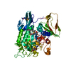


 Mass: 92.094 Da / Num. of mol.: 1 / Source method: obtained synthetically / Formula: C3H8O3
 Mass: 18.015 Da / Num. of mol.: 480 / Source method: isolated from a natural source / Formula: H2O
 Sample preparation
 / Beamline: 8.2.1 / Wavelength: 0.918401,0.979261,0.979152
 Processing