 Movie
Movie Controller

[English] 日本語
 Yorodumi
- PDB-4ny7: Bond length analysis of the PqqC Y175F mutant structure shows evi... -
+ Open data

- Basic information
| Entry | Database: PDB / ID: 4ny7 | ||||||
|---|---|---|---|---|---|---|---|
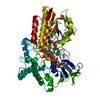
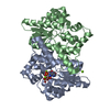
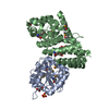
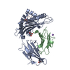
| Title | Bond length analysis of the PqqC Y175F mutant structure shows evidence for bound PQQ in the reduced form | ||||||
 Components Components | Pyrroloquinoline-quinone synthase | ||||||
 Keywords Keywords | OXIDOREDUCTASE / all helical | ||||||
| Function / homology |  Function and homology information Function and homology informationpyrroloquinoline-quinone synthase / pyrroloquinoline-quinone synthase activity / pyrroloquinoline quinone biosynthetic process Similarity search - Function | ||||||
| Biological species |  Klebsiella pneumoniae subsp. pneumoniae (bacteria) Klebsiella pneumoniae subsp. pneumoniae (bacteria) | ||||||
| Method |  X-RAY DIFFRACTION / SYNCHROTRON / MOLECULAR REPLACEMENT / Resolution: 1.44 Å X-RAY DIFFRACTION / SYNCHROTRON / MOLECULAR REPLACEMENT / Resolution: 1.44 Å | ||||||
 Authors Authors | Fisher, S.J. / Puehringer, S. | ||||||
 Citation Citation | Journal: To be Published Title: Bond length analysis of the PqqC Y175F mutant structure shows evidence for bound PQQ in the reduced form Authors: Fisher, S.J. / Puehringer, S. | ||||||
| History |
|
- Structure visualization
| Structure viewer | Molecule: MolmilJmol/JSmol |
|---|
- Downloads & links
-Download
| PDBx/mmCIF format | 4ny7.cif.gz | 264.1 KB | Display | PDBx/mmCIF format |
|---|---|---|---|---|
| PDB format | pdb4ny7.ent.gz | 210.9 KB | Display | PDB format |
| PDBx/mmJSON format | 4ny7.json.gz | Tree view | PDBx/mmJSON format | |
| Others |  Other downloads Other downloads |
-Validation report
| Arichive directory | https://data.pdbj.org/pub/pdb/validation_reports/ny/4ny7ftp://data.pdbj.org/pub/pdb/validation_reports/ny/4ny7 | HTTPS FTP |
|---|
-Related structure data
| Related structure data |  3hlxS S: Starting model for refinement |
|---|---|
| Similar structure data |


-Links
 PDBj
PDBj
- Assembly
| Deposited unit | 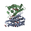
| ||||||||
|---|---|---|---|---|---|---|---|---|---|
| 1 |
| ||||||||
| Unit cell |
|
-Components
| #1: Protein | Mass: 29986.918 Da / Num. of mol.: 2 / Mutation: Y175F Source method: isolated from a genetically manipulated source Source: (gene. exp.) Klebsiella pneumoniae subsp. pneumoniae (bacteria)Strain: ATCC 700721 / MGH 78578 / Gene: pqqC, KPN78578_17810, KPN_01811 / Production host: References: UniProt: A6T9H1, pyrroloquinoline-quinone synthase #2: Chemical |   Mass: 330.206 Da / Num. of mol.: 2 / Source method: obtained synthetically / Formula: C14H6N2O8 Mass: 330.206 Da / Num. of mol.: 2 / Source method: obtained synthetically / Formula: C14H6N2O8#3: Chemical | ChemComp-GOL /   Mass: 92.094 Da / Num. of mol.: 10 / Source method: obtained synthetically / Formula: C3H8O3 Mass: 92.094 Da / Num. of mol.: 10 / Source method: obtained synthetically / Formula: C3H8O3#4: Chemical |   Mass: 35.453 Da / Num. of mol.: 2 / Source method: obtained synthetically / Formula: Cl Mass: 35.453 Da / Num. of mol.: 2 / Source method: obtained synthetically / Formula: Cl#5: Water | ChemComp-HOH / |  Mass: 18.015 Da / Num. of mol.: 568 / Source method: isolated from a natural source / Formula: H2O Mass: 18.015 Da / Num. of mol.: 568 / Source method: isolated from a natural source / Formula: H2O |
|---|
-Experimental details
-Experiment
| Experiment | Method: X-RAY DIFFRACTION / Number of used crystals: 1 |
|---|
- Sample preparation
| Crystal | Density Matthews: 2.32 Å3/Da / Density % sol: 46.95 % |
|---|---|
| Crystal grow | Temperature: 298 K / Method: vapor diffusion, sitting drop / pH: 7.8 Details: 0.2M ammonium sulfate, 0.1M Bis-Tris pH 6.5, 25% w/v poly- ethylene glycol 3,350, VAPOR DIFFUSION, SITTING DROP, temperature 298K |
-Data collection
| Diffraction | Mean temperature: 100 K |
|---|---|
| Diffraction source | Source: SYNCHROTRON / Site: ESRF  / Beamline: ID14-1 / Wavelength: 0.934 Å / Beamline: ID14-1 / Wavelength: 0.934 Å |
| Detector | Type: ADSC QUANTUM 210 / Detector: CCD |
| Radiation | Monochromator: Diamond (111), Ge(220) / Protocol: SINGLE WAVELENGTH / Monochromatic (M) / Laue (L): M / Scattering type: x-ray |
| Radiation wavelength | Wavelength: 0.934 Å / Relative weight: 1 |
| Reflection | Resolution: 1.43→37.72 Å / Num. all: 102022 / Num. obs: 100186 / % possible obs: 98.2 % / Observed criterion σ(F): 2 / Observed criterion σ(I): 2 / Redundancy: 6.9 % / Rmerge(I) obs: 0.056 / Net I/σ(I): 21 |
| Reflection shell | Resolution: 1.43→1.51 Å / Redundancy: 5.5 % / Rmerge(I) obs: 0.374 / Mean I/σ(I) obs: 4.3 / % possible all: 0.903 |
- Processing
| Software |
| |||||||||||||||||||||||||||||||||
|---|---|---|---|---|---|---|---|---|---|---|---|---|---|---|---|---|---|---|---|---|---|---|---|---|---|---|---|---|---|---|---|---|---|---|
| Refinement | Method to determine structure: MOLECULAR REPLACEMENT Starting model: pdb entry 3HLX Resolution: 1.44→10 Å / Stereochemistry target values: Engh & Huber
| |||||||||||||||||||||||||||||||||
| Refinement step | Cycle: LAST / Resolution: 1.44→10 Å
| |||||||||||||||||||||||||||||||||
| Refine LS restraints |
|
