 Movie
Movie Controller

[English] 日本語
 Yorodumi
- PDB-1ih8: NH3-dependent NAD+ Synthetase from Bacillus subtilis Complexed wi... -
+ Open data

- Basic information
| Entry | Database: PDB / ID: 1ih8 | ||||||
|---|---|---|---|---|---|---|---|
| Title | NH3-dependent NAD+ Synthetase from Bacillus subtilis Complexed with AMP-CPP and Mg2+ ions. | ||||||
 Components Components | NH(3)-DEPENDENT NAD(+) synthetase | ||||||
 Keywords Keywords | LIGASE / amidotransferase / ATP pyrophosphatase / active-site loops | ||||||
| Function / homology |  Function and homology information Function and homology informationNAD+ synthase / NAD+ synthase activity / NAD+ synthase (glutamine-hydrolyzing) activity / glutaminase activity / NAD+ biosynthetic process / sporulation resulting in formation of a cellular spore / ATP binding / metal ion binding / cytoplasm Similarity search - Function | ||||||
| Biological species |  | ||||||
| Method |  X-RAY DIFFRACTION / FOURIER SYNTHESIS / Resolution: 1.9 Å X-RAY DIFFRACTION / FOURIER SYNTHESIS / Resolution: 1.9 Å | ||||||
 Authors Authors | Devedjiev, Y. / Symersky, J. / Singh, R. / Jedrzejas, M. / Brouillette, C. / Brouillette, W. / Muccio, D. / Chattopadhyay, D. / DeLucas, L. | ||||||
 Citation Citation | Journal: Acta Crystallogr.,Sect.D / Year: 2001 Title: Stabilization of active-site loops in NH3-dependent NAD+ synthetase from Bacillus subtilis. Authors: Devedjiev, Y. / Symersky, J. / Singh, R. / Jedrzejas, M. / Brouillette, C. / Brouillette, W. / Muccio, D. / Chattopadhyay, D. / DeLucas, L. | ||||||
| History |
|
- Structure visualization
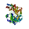
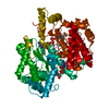
| Structure viewer | Molecule: MolmilJmol/JSmol |
|---|
- Downloads & links
-Download
| PDBx/mmCIF format | 1ih8.cif.gz | 130.4 KB | Display | PDBx/mmCIF format |
|---|---|---|---|---|
| PDB format | pdb1ih8.ent.gz | 98.4 KB | Display | PDB format |
| PDBx/mmJSON format | 1ih8.json.gz | Tree view | PDBx/mmJSON format | |
| Others |  Other downloads Other downloads |
-Validation report
| Arichive directory | https://data.pdbj.org/pub/pdb/validation_reports/ih/1ih8ftp://data.pdbj.org/pub/pdb/validation_reports/ih/1ih8 | HTTPS FTP |
|---|
-Related structure data
| Related structure data |  1ee1C  1fydC  1ifxC  1nsyS S: Starting model for refinement C: citing same article ( |
|---|---|
| Similar structure data |


-Links
 PDBj
PDBj


- Assembly
| Deposited unit | 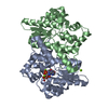
| ||||||||
|---|---|---|---|---|---|---|---|---|---|
| 1 |
| ||||||||
| Unit cell |
| ||||||||
| Details | The biological assembly is a homodimer consisting of chains A and B in the same asymmetric unit. |
-Components
| #1: Protein | Mass: 30303.994 Da / Num. of mol.: 2 Source method: isolated from a genetically manipulated source Source: (gene. exp.) References: UniProt: P08164, NAD+ synthase (glutamine-hydrolysing) #2: Chemical |   Mass: 24.305 Da / Num. of mol.: 3 / Source method: obtained synthetically / Formula: Mg Mass: 24.305 Da / Num. of mol.: 3 / Source method: obtained synthetically / Formula: Mg#3: Chemical | ChemComp-APC / |   Mass: 505.208 Da / Num. of mol.: 1 / Source method: obtained synthetically / Formula: C11H18N5O12P3 / Comment: AMP-CPP, energy-carrying molecule analogue*YM Mass: 505.208 Da / Num. of mol.: 1 / Source method: obtained synthetically / Formula: C11H18N5O12P3 / Comment: AMP-CPP, energy-carrying molecule analogue*YM#4: Water | ChemComp-HOH / |  Mass: 18.015 Da / Num. of mol.: 590 / Source method: isolated from a natural source / Formula: H2O Mass: 18.015 Da / Num. of mol.: 590 / Source method: isolated from a natural source / Formula: H2O |
|---|
-Experimental details
-Experiment
| Experiment | Method: X-RAY DIFFRACTION / Number of used crystals: 1 |
|---|
- Sample preparation
| Crystal | Density Matthews: 2.1 Å3/Da / Density % sol: 39 % | ||||||||||||||||||||||||||||||||||||||||||
|---|---|---|---|---|---|---|---|---|---|---|---|---|---|---|---|---|---|---|---|---|---|---|---|---|---|---|---|---|---|---|---|---|---|---|---|---|---|---|---|---|---|---|---|
| Crystal grow | Temperature: 295 K / Method: vapor diffusion, hanging drop / pH: 7.5 Details: VAPOR DIFFUSION IN MICROSEEDED HANGING DROPS. 20-26% PEG400, 0.05 M HEPES SODIUM SALT, PH=7.5, 0.1 M MAGNESIUM CHLORIDE, 0.002 M AMP-CPP., VAPOR DIFFUSION, HANGING DROP, temperature 295K | ||||||||||||||||||||||||||||||||||||||||||
| Crystal grow | *PLUS Details: used microseeding | ||||||||||||||||||||||||||||||||||||||||||
| Components of the solutions | *PLUS
|
-Data collection
| Diffraction | Mean temperature: 120 K |
|---|---|
| Diffraction source | Source: ROTATING ANODE / Type: RIGAKU RU200 / Wavelength: 1.5418 Å |
| Detector | Type: MACSCIENCE DIP100 / Detector: IMAGE PLATE / Date: Jun 27, 2000 / Details: mirrors |
| Radiation | Monochromator: Ni filter / Protocol: SINGLE WAVELENGTH / Monochromatic (M) / Laue (L): M / Scattering type: x-ray |
| Radiation wavelength | Wavelength: 1.5418 Å / Relative weight: 1 |
| Reflection | Resolution: 1.9→30 Å / Num. all: 112884 / Num. obs: 38796 / % possible obs: 98.8 % / Observed criterion σ(F): 0 / Observed criterion σ(I): -10 / Redundancy: 2.9 % / Biso Wilson estimate: 10.8 Å2 / Rmerge(I) obs: 0.048 / Net I/σ(I): 20.4 |
| Reflection shell | Resolution: 1.9→1.97 Å / Redundancy: 2.5 % / Rmerge(I) obs: 0.154 / Mean I/σ(I) obs: 7.6 / % possible all: 98.4 |
| Reflection | *PLUS Num. measured all: 112884 |
| Reflection shell | *PLUS % possible obs: 98.8 % |
- Processing
| Software |
| ||||||||||||||||||||||||||||||||||||
|---|---|---|---|---|---|---|---|---|---|---|---|---|---|---|---|---|---|---|---|---|---|---|---|---|---|---|---|---|---|---|---|---|---|---|---|---|---|
| Refinement | Method to determine structure: FOURIER SYNTHESIS Starting model: pdb entry 1NSY Resolution: 1.9→29.83 Å / Rfactor Rfree error: 0.005 / Data cutoff high absF: 346000.4 / Data cutoff high rms absF: 10000 / Data cutoff low absF: 0 / Isotropic thermal model: RESTRAINED / Cross valid method: THROUGHOUT / σ(F): 2 / σ(I): 1.36 / Stereochemistry target values: Engh & Huber Details: Subunit A has all 271 residues and methyleneadenosine triphosphate. In the subunit B, residues 1083-1086, 1205-1224, and methyleneadenosine triphosphate are disordered and not included in the model.
| ||||||||||||||||||||||||||||||||||||
| Solvent computation | Solvent model: FLAT MODEL / Bsol: 48.29 Å2 / ksol: 0.371 e/Å3 | ||||||||||||||||||||||||||||||||||||
| Displacement parameters | Biso mean: 15.4 Å2
| ||||||||||||||||||||||||||||||||||||
| Refine analyze |
| ||||||||||||||||||||||||||||||||||||
| Refinement step | Cycle: LAST / Resolution: 1.9→29.83 Å
| ||||||||||||||||||||||||||||||||||||
| Refine LS restraints |
| ||||||||||||||||||||||||||||||||||||
| LS refinement shell | Resolution: 1.9→1.97 Å / Rfactor Rfree error: 0.017 / Total num. of bins used: 10
| ||||||||||||||||||||||||||||||||||||
| Xplor file |
| ||||||||||||||||||||||||||||||||||||
| Software | *PLUS Name: CNS / Version: 1 / Classification: refinement | ||||||||||||||||||||||||||||||||||||
| Refinement | *PLUS σ(F): 2 / % reflection Rfree: 5 % | ||||||||||||||||||||||||||||||||||||
| Solvent computation | *PLUS | ||||||||||||||||||||||||||||||||||||
| Displacement parameters | *PLUS Biso mean: 15.4 Å2 | ||||||||||||||||||||||||||||||||||||
| Refine LS restraints | *PLUS
| ||||||||||||||||||||||||||||||||||||
| LS refinement shell | *PLUS Rfactor Rfree: 0.244 / % reflection Rfree: 5.5 % / Rfactor Rwork: 0.175 |
