Resolution: 1.449→1.48 Å / Redundancy: 6.6 % / Rmerge(I) obs: 0.249 / Mean I/σ(I) obs: 10.07 / Num. unique all: 1475 / Rsym value: 0.212 / % possible all: 98.7
-
Processing
Software
Name
Version
Classification
HKL-3000
datacollection
HKL-3000
phasing
REFMAC
5.7.0029
refinement
HKL-3000
datareduction
HKL-3000
datascaling
Refinement
Method to determine structure: SAD / Resolution: 1.449→24.52 Å / Cor.coef. Fo:Fc: 0.963 / Cor.coef. Fo:Fc free: 0.947 / SU B: 1.208 / SU ML: 0.047 / Cross valid method: THROUGHOUT / ESU R: 0.07 / ESU R Free: 0.075 / Stereochemistry target values: MAXIMUM LIKELIHOOD / Details: HYDROGENS HAVE BEEN ADDED IN THE RIDING POSITIONS
Rfactor
Num. reflection
% reflection
Selection details
Rfree
0.21678
1540
5 %
RANDOM
Rwork
0.17649
-
-
-
all
0.17845
29002
-
-
obs
0.17845
29002
99.66 %
-
Solvent computation
Ion probe radii: 0.8 Å / Shrinkage radii: 0.8 Å / VDW probe radii: 1.2 Å / Solvent model: MASK
 Movie
Movie Controller


 Open data
 Basic information
 Components
 Keywords
 Function and homology information
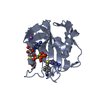
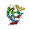
 Streptomyces lividans (bacteria)
 X-RAY DIFFRACTION /
  Authors
 Citation
 Structure visualization
 Downloads & links
 Other downloads
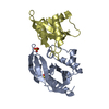


 PDBj
PDBj


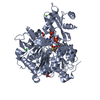
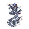
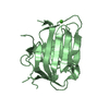
 Assembly
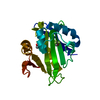
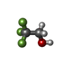
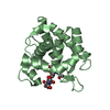
 Mass: 100.040 Da / Num. of mol.: 1 / Source method: obtained synthetically / Formula: C2H3F3O
 Mass: 92.094 Da / Num. of mol.: 1 / Source method: obtained synthetically / Formula: C3H8O3
 Mass: 18.015 Da / Num. of mol.: 185 / Source method: isolated from a natural source / Formula: H2O
 Sample preparation
 / Beamline: 19-ID / Wavelength: 0.97936 Å
 Processing