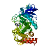
 Movie
Movie Controller

[English] 日本語
 Yorodumi
- PDB-4u5y: Crystal Structure of the complex between the GNAT domain of S. li... -
+ Open data

- Basic information
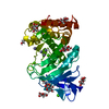
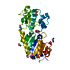
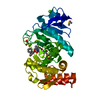
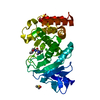
| Entry | Database: PDB / ID: 4u5y | |||||||||
|---|---|---|---|---|---|---|---|---|---|---|
| Title | Crystal Structure of the complex between the GNAT domain of S. lividans PAT and the acetyl-CoA synthetase C-terminal domain of S. enterica | |||||||||
 Components Components |
| |||||||||
 Keywords Keywords | LIGASE / GNAT / COMPLEX / ACETYLATION / PAT | |||||||||
| Function / homology |  Function and homology information Function and homology informationN-acetyltransferase activity / acyltransferase activity, transferring groups other than amino-acyl groups Similarity search - Function | |||||||||
| Biological species |  Streptomyces lividans TK24 (bacteria)Salmonella enterica subsp. enterica serovar Paratyphi B str. ATCC 10719 (bacteria) Streptomyces lividans TK24 (bacteria)Salmonella enterica subsp. enterica serovar Paratyphi B str. ATCC 10719 (bacteria) | |||||||||
| Method |  X-RAY DIFFRACTION / SYNCHROTRON / MOLECULAR REPLACEMENT / Resolution: 1.926 Å X-RAY DIFFRACTION / SYNCHROTRON / MOLECULAR REPLACEMENT / Resolution: 1.926 Å | |||||||||
 Authors Authors | Taylor, K.C. / Tucker, A.C. / Rank, K.C. / Escalante-Semerena, J.C. / Rayment, I. | |||||||||
| Funding support |  United States, 2items United States, 2items
| |||||||||
 Citation Citation | Journal: J.Biol.Chem. / Year: 2014 Title: Insights into the specificity of lysine acetyltransferases. Authors: Tucker, A.C. / Taylor, K.C. / Rank, K.C. / Rayment, I. / Escalante-Semerena, J.C. | |||||||||
| History |
|
- Structure visualization
| Structure viewer | Molecule: MolmilJmol/JSmol |
|---|
- Downloads & links
-Download
| PDBx/mmCIF format | 4u5y.cif.gz | 75.1 KB | Display | PDBx/mmCIF format |
|---|---|---|---|---|
| PDB format | pdb4u5y.ent.gz | 53.3 KB | Display | PDB format |
| PDBx/mmJSON format | 4u5y.json.gz | Tree view | PDBx/mmJSON format | |
| Others |  Other downloads Other downloads |
-Validation report
| Arichive directory | https://data.pdbj.org/pub/pdb/validation_reports/u5/4u5yftp://data.pdbj.org/pub/pdb/validation_reports/u5/4u5y | HTTPS FTP |
|---|
-Related structure data
| Related structure data | 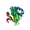 4nxySC 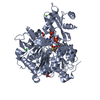 1pg4S S: Starting model for refinement C: citing same article ( |
|---|---|
| Similar structure data |


-Links
 PDBj
PDBj


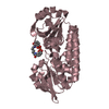
- Assembly
| Deposited unit | 
| ||||||||
|---|---|---|---|---|---|---|---|---|---|
| 1 |
| ||||||||
| Unit cell |
| ||||||||
| Components on special symmetry positions |
|
-Components
| #1: Protein | Mass: 21669.332 Da / Num. of mol.: 1 / Fragment: UNP residues 1-194 / Mutation: S73C Source method: isolated from a genetically manipulated source Source: (gene. exp.) Streptomyces lividans TK24 (bacteria) / Gene: SSPG_01886 / Production host: | ||
|---|---|---|---|
| #2: Protein | Mass: 14535.637 Da / Num. of mol.: 1 / Fragment: UNP residues 518-652 / Mutation: H567C Source method: isolated from a genetically manipulated source Source: (gene. exp.) Salmonella enterica subsp. enterica serovar Paratyphi B str. ATCC 10719 (bacteria)Gene: acs, SEEPB719_19728 / Production host: | ||
| #3: Chemical |   Mass: 96.063 Da / Num. of mol.: 2 / Source method: obtained synthetically / Formula: SO4 Mass: 96.063 Da / Num. of mol.: 2 / Source method: obtained synthetically / Formula: SO4#4: Water | ChemComp-HOH / |  Mass: 18.015 Da / Num. of mol.: 185 / Source method: isolated from a natural source / Formula: H2O Mass: 18.015 Da / Num. of mol.: 185 / Source method: isolated from a natural source / Formula: H2O |
-Experimental details
-Experiment
| Experiment | Method: X-RAY DIFFRACTION / Number of used crystals: 1 |
|---|
- Sample preparation
| Crystal | Density Matthews: 1.99 Å3/Da / Density % sol: 38.13 % |
|---|---|
| Crystal grow | Temperature: 293 K / Method: vapor diffusion, hanging drop / pH: 6.5 Details: 11.2% (w/v) polyethylene glycol 8000, 100 mM 1,4-piperazinediethanesulfonic acid pH 6.5, 120 mM Li2SO4 |
-Data collection
| Diffraction | Mean temperature: 100 K |
|---|---|
| Diffraction source | Source: SYNCHROTRON / Site: APS / Beamline: 19-BM / Wavelength: 0.978 Å |
| Detector | Type: ADSC QUANTUM 210r / Detector: CCD / Date: Apr 14, 2014 |
| Radiation | Monochromator: Rosenbaum-Rock double-crystal monochromator: Water cooled; sagitally focusing 2nd crystal, Rosenbaum-Rock vertical focusing mirror Protocol: SINGLE WAVELENGTH / Monochromatic (M) / Laue (L): M / Scattering type: x-ray |
| Radiation wavelength | Wavelength: 0.978 Å / Relative weight: 1 |
| Reflection | Resolution: 1.9→50 Å / Num. obs: 22055 / % possible obs: 99.7 % / Redundancy: 10.6 % / Rmerge(I) obs: 0.067 / Net I/av σ(I): 30.5 / Net I/σ(I): 24.3 |
| Reflection shell | Resolution: 1.92→1.95 Å / Redundancy: 7.3 % / Rmerge(I) obs: 0.33 / Mean I/σ(I) obs: 4.9 / % possible all: 98.4 |
- Processing
| Software | Name: PHENIX / Version: (phenix.refine: 1.8.4_1496) / Classification: refinement | ||||||||||||||||||||||||||||||||||||||||||||||||||||||||
|---|---|---|---|---|---|---|---|---|---|---|---|---|---|---|---|---|---|---|---|---|---|---|---|---|---|---|---|---|---|---|---|---|---|---|---|---|---|---|---|---|---|---|---|---|---|---|---|---|---|---|---|---|---|---|---|---|---|
| Refinement | Method to determine structure: MOLECULAR REPLACEMENT Starting model: 4NXY, 1PG4 Resolution: 1.926→21.632 Å / SU ML: 0.25 / Cross valid method: FREE R-VALUE / σ(F): 1.36 / Phase error: 22.63 / Stereochemistry target values: ML
| ||||||||||||||||||||||||||||||||||||||||||||||||||||||||
| Solvent computation | Shrinkage radii: 0.9 Å / VDW probe radii: 1.11 Å / Solvent model: FLAT BULK SOLVENT MODEL | ||||||||||||||||||||||||||||||||||||||||||||||||||||||||
| Displacement parameters | Biso mean: 18.8 Å2 | ||||||||||||||||||||||||||||||||||||||||||||||||||||||||
| Refinement step | Cycle: LAST / Resolution: 1.926→21.632 Å
| ||||||||||||||||||||||||||||||||||||||||||||||||||||||||
| Refine LS restraints |
| ||||||||||||||||||||||||||||||||||||||||||||||||||||||||
| LS refinement shell | Refine-ID: X-RAY DIFFRACTION
|
