[English] 日本語
Yorodumi
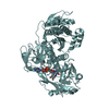
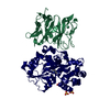
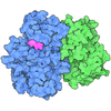
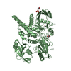
- PDB-4nwg: Crystal structure of the tyrosine phosphatase SHP-2 with E139D mu... -

+
Open data


ID or keywords:

Loading...

-
Basic information

Entry
Database: PDB / ID: 4nwg
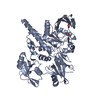
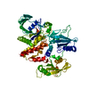
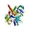
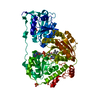
TitleCrystal structure of the tyrosine phosphatase SHP-2 with E139D mutation
ComponentsTyrosine-protein phosphatase non-receptor type 11
KeywordsHYDROLASE
Function / homology
Function and homology information


atrioventricular canal development / genitalia development / STAT5 Activation / Co-inhibition by BTLA / Netrin mediated repulsion signals / negative regulation of neutrophil activation / negative regulation of chondrocyte differentiation / positive regulation of lipopolysaccharide-mediated signaling pathway / face morphogenesis / Interleukin-37 signaling ...atrioventricular canal development / genitalia development / STAT5 Activation / Co-inhibition by BTLA / Netrin mediated repulsion signals / negative regulation of neutrophil activation / negative regulation of chondrocyte differentiation / positive regulation of lipopolysaccharide-mediated signaling pathway / face morphogenesis / Interleukin-37 signaling / positive regulation of ossification / Signaling by Leptin / negative regulation of cell adhesion mediated by integrin / MET activates PTPN11 / Regulation of RUNX1 Expression and Activity / Signal regulatory protein family interactions / ERBB signaling pathway / Interleukin-20 family signaling / Interleukin-6 signaling / Co-inhibition by CTLA4 / PI-3K cascade:FGFR3 / STAT5 activation downstream of FLT3 ITD mutants / Platelet sensitization by LDL / negative regulation of T cell activation / inner ear development / fibroblast growth factor receptor signaling pathway / PI-3K cascade:FGFR2 / PI-3K cascade:FGFR4 / peptide hormone receptor binding / negative regulation of type I interferon production / MAPK3 (ERK1) activation / PI-3K cascade:FGFR1 / regulation of type I interferon-mediated signaling pathway / MAPK1 (ERK2) activation / Prolactin receptor signaling / PECAM1 interactions / non-membrane spanning protein tyrosine phosphatase activity / peptidyl-tyrosine dephosphorylation / Regulation of IFNA/IFNB signaling / positive regulation of intracellular signal transduction / RET signaling / Interleukin-3, Interleukin-5 and GM-CSF signaling / Co-inhibition by PD-1 / PI3K Cascade / ephrin receptor signaling pathway / positive regulation of insulin receptor signaling pathway / regulation of protein-containing complex assembly / negative regulation of T cell receptor signaling pathway / negative regulation of T cell proliferation / Regulation of IFNG signaling / GAB1 signalosome / T cell costimulation / Activated NTRK2 signals through FRS2 and FRS3 / GPVI-mediated activation cascade / Signaling by CSF3 (G-CSF) / FRS-mediated FGFR3 signaling / Signaling by FLT3 ITD and TKD mutants / phosphotyrosine residue binding / FRS-mediated FGFR2 signaling / FRS-mediated FGFR4 signaling / phosphoprotein phosphatase activity / protein-tyrosine-phosphatase / FRS-mediated FGFR1 signaling / Tie2 Signaling / positive regulation of D-glucose import across plasma membrane / FLT3 Signaling / protein tyrosine phosphatase activity / cell adhesion molecule binding / positive regulation of interferon-beta production / Downstream signal transduction / cellular response to epidermal growth factor stimulus / protein tyrosine kinase binding / insulin receptor binding / Activation of IRF3, IRF7 mediated by TBK1, IKKε (IKBKE) / brain development / Negative regulation of FGFR3 signaling / cellular response to mechanical stimulus / Negative regulation of FGFR2 signaling / Negative regulation of FGFR4 signaling / Negative regulation of FGFR1 signaling / Signaling by SCF-KIT / Spry regulation of FGF signaling / receptor tyrosine kinase binding / vasodilation / epidermal growth factor receptor signaling pathway / cytokine-mediated signaling pathway / Constitutive Signaling by Aberrant PI3K in Cancer / heart development / Signaling by CSF1 (M-CSF) in myeloid cells / Interferon alpha/beta signaling / positive regulation of tumor necrosis factor production / PIP3 activates AKT signaling / PI5P, PP2A and IER3 Regulate PI3K/AKT Signaling / signaling receptor complex adaptor activity / positive regulation of phosphatidylinositol 3-kinase/protein kinase B signal transduction / molecular adaptor activity / positive regulation of ERK1 and ERK2 cascade / cadherin binding / focal adhesion / nucleolus
Similarity search - Function
Protein-tyrosine phosphatase, non-receptor type-6, -11 / SH2 domain / SHC Adaptor Protein / Protein tyrosine phosphatase superfamily / Protein-Tyrosine Phosphatase; Chain A / Protein tyrosine phosphatase, catalytic domain / PTP type protein phosphatase domain profile. / Protein-tyrosine phosphatase / Tyrosine-specific protein phosphatase, PTPase domain / Protein-tyrosine phosphatase, catalytic ...Protein-tyrosine phosphatase, non-receptor type-6, -11 / SH2 domain / SHC Adaptor Protein / Protein tyrosine phosphatase superfamily / Protein-Tyrosine Phosphatase; Chain A / Protein tyrosine phosphatase, catalytic domain / PTP type protein phosphatase domain profile. / Protein-tyrosine phosphatase / Tyrosine-specific protein phosphatase, PTPase domain / Protein-tyrosine phosphatase, catalytic / Protein tyrosine phosphatase, catalytic domain motif / Tyrosine specific protein phosphatases active site. / Protein-tyrosine phosphatase, active site / Tyrosine specific protein phosphatases domain profile. / Tyrosine-specific protein phosphatases domain / Protein-tyrosine phosphatase-like / SH2 domain / Src homology 2 (SH2) domain profile. / Src homology 2 domains / SH2 domain / SH2 domain superfamily / Alpha-Beta Complex / 2-Layer Sandwich / Alpha Beta
Similarity search - Domain/homology
DI(HYDROXYETHYL)ETHER / Tyrosine-protein phosphatase non-receptor type 11
Similarity search - Component
Biological speciesHomo sapiens (human)
MethodX-RAY DIFFRACTION / SYNCHROTRON / MOLECULAR REPLACEMENT / Resolution: 2.45 Å
AuthorsQiu, W. / Lin, A. / Hutchinson, A. / Romanov, V. / Ruzanov, M. / Thompson, C. / Lam, K. / Kisselman, G. / Battalie, K. / Chirgadze, N.Y.
CitationJournal: To be Published
Title: Crystal structure of the tyrosine phosphatase SHP-2 with E139D mutation
Authors: Qiu, W. / Lin, A. / Hutchinson, A. / Romanov, V. / Ruzanov, M. / Thompson, C. / Lam, K. / Kisselman, G. / Battalie, K. / Chirgadze, N.Y.
History
DepositionDec 6, 2013Deposition site: RCSB / Processing site: RCSB
Revision 1.0Dec 10, 2014Provider: repository / Type: Initial release
Revision 1.1Feb 28, 2024Group: Data collection / Database references / Derived calculations
Category: chem_comp_atom / chem_comp_bond ...chem_comp_atom / chem_comp_bond / database_2 / struct_ref_seq_dif / struct_site
Item: _database_2.pdbx_DOI / _database_2.pdbx_database_accession ..._database_2.pdbx_DOI / _database_2.pdbx_database_accession / _struct_ref_seq_dif.details / _struct_site.pdbx_auth_asym_id / _struct_site.pdbx_auth_comp_id / _struct_site.pdbx_auth_seq_id

-
Structure visualization

Structure viewerMolecule:
MolmilJmol/JSmol

Downloads & links

-
Assembly

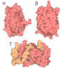
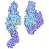
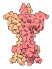
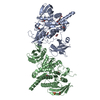
Deposited unit
A: Tyrosine-protein phosphatase non-receptor type 11
B: Tyrosine-protein phosphatase non-receptor type 11
hetero molecules


Theoretical massNumber of molelcules
Total (without water)124,87315
Polymers123,9882
Non-polymers88513
Water6,521362
1
A: Tyrosine-protein phosphatase non-receptor type 11
hetero molecules


Theoretical massNumber of molelcules
Total (without water)62,5359
Polymers61,9941
Non-polymers5418
Water181
TypeNameSymmetry operationNumber
identity operation1_555x,y,z1
MethodPISA
2
B: Tyrosine-protein phosphatase non-receptor type 11
hetero molecules


Theoretical massNumber of molelcules
Total (without water)62,3386
Polymers61,9941
Non-polymers3445
Water181
TypeNameSymmetry operationNumber
identity operation1_555x,y,z1
MethodPISA
Unit cell
Length a, b, c (Å)56.320, 212.420, 92.160
Angle α, β, γ (deg.)90.00, 90.00, 90.00
Int Tables number19
Space group name H-MP212121

-
Components

#1: Protein Tyrosine-protein phosphatase non-receptor type 11 / Protein-tyrosine phosphatase 1D / PTP-1D / Protein-tyrosine phosphatase 2C / PTP-2C / SH-PTP2 / SHP- ...Protein-tyrosine phosphatase 1D / PTP-1D / Protein-tyrosine phosphatase 2C / PTP-2C / SH-PTP2 / SHP-2 / Shp2 / SH-PTP3


Mass: 61993.957 Da / Num. of mol.: 2 / Fragment: UNP residues 1-543 / Mutation: E139D
Source method: isolated from a genetically manipulated source
Source: (gene. exp.) Homo sapiens (human) / Gene: PTPN11, PTP2C, SHPTP2 / Production host: Escherichia coli (E. coli) / References: UniProt: Q06124, protein-tyrosine-phosphatase
#2: Chemical
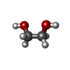
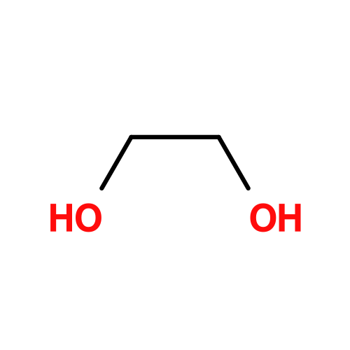
ChemComp-EDO / 1,2-ETHANEDIOL / ETHYLENE GLYCOL


Mass: 62.068 Da / Num. of mol.: 11 / Source method: obtained synthetically / Formula: C2H6O2
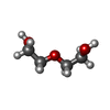
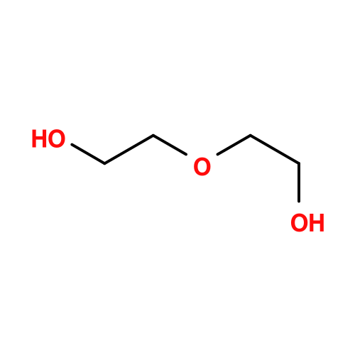
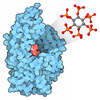
#3: Chemical ChemComp-PEG / DI(HYDROXYETHYL)ETHER


Mass: 106.120 Da / Num. of mol.: 1 / Source method: obtained synthetically / Formula: C4H10O3
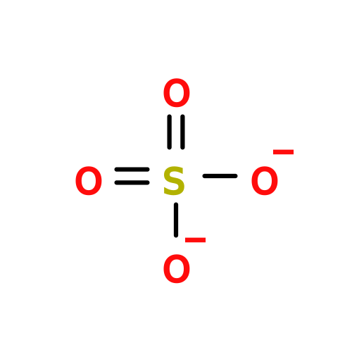
#4: Chemical ChemComp-SO4 / SULFATE ION


Mass: 96.063 Da / Num. of mol.: 1 / Source method: obtained synthetically / Formula: SO4

#5: Water ChemComp-HOH / water


Mass: 18.015 Da / Num. of mol.: 362 / Source method: isolated from a natural source / Formula: H2O

-
Experimental details

-
Experiment

ExperimentMethod: X-RAY DIFFRACTION

-
Sample preparation

CrystalDensity Matthews: 2.22 Å3/Da / Density % sol: 44.67 %
Crystal growTemperature: 293 K / Method: vapor diffusion / pH: 8
Details: 14.5% PEG4000, 0.1M Tris buffer, 0.02M DDT, pH 8.0, VAPOR DIFFUSION, temperature 293K

-
Data collection

DiffractionMean temperature: 100 K
Diffraction sourceSource: SYNCHROTRON / Site: APS / Beamline: 17-ID / Wavelength: 1 Å
DetectorType: PSI PILATUS 6M / Detector: PIXEL / Date: Oct 28, 2012
RadiationMonochromator: Si 111 / Protocol: SINGLE WAVELENGTH / Monochromatic (M) / Laue (L): M / Scattering type: x-ray
Radiation wavelengthWavelength: 1 Å / Relative weight: 1
ReflectionResolution: 2.45→100 Å / Num. all: 41754 / Num. obs: 41625 / % possible obs: 99.7 % / Observed criterion σ(F): 0 / Observed criterion σ(I): 0 / Redundancy: 6.51 % / Biso Wilson estimate: 49.87 Å2 / Rmerge(I) obs: 0.1137 / Rsym value: 0.0682 / Net I/σ(I): 9.19
Reflection shellResolution: 2.45→2.55 Å / Redundancy: 6.28 % / Rmerge(I) obs: 0.5712 / Mean I/σ(I) obs: 2.31 / Num. unique all: 4590 / Rsym value: 0.4437 / % possible all: 98.1

-
Processing

Software
NameVersionClassification
JDirectordata collection
PHASERphasing
BUSTER2.10.0refinement
XDSdata reduction
XDSdata scaling
RefinementMethod to determine structure: MOLECULAR REPLACEMENT / Resolution: 2.45→38.64 Å / Cor.coef. Fo:Fc: 0.9128 / Cor.coef. Fo:Fc free: 0.867 / SU R Cruickshank DPI: 0.718 / Cross valid method: THROUGHOUT / σ(F): 0 / Stereochemistry target values: Engh & Huber
RfactorNum. reflection% reflectionSelection details
Rfree0.3258 1070 2.58 %RANDOM
Rwork0.2804 ---
obs0.2815 41540 99.9 %-
all-41625 --
Displacement parametersBiso mean: 48.56 Å2
Baniso -1Baniso -2Baniso -3
1-2.1143 Å20 Å20 Å2
2--7.771 Å20 Å2
3----9.8853 Å2
Refine analyzeLuzzati coordinate error obs: 0.422 Å
Refinement stepCycle: LAST / Resolution: 2.45→38.64 Å
ProteinNucleic acidLigandSolventTotal
Num. atoms8021 0 56 362 8439
Refine LS restraints
Refine-IDTypeDev idealNumberRestraint functionWeight
X-RAY DIFFRACTIONt_bond_d0.0078245HARMONIC2
X-RAY DIFFRACTIONt_angle_deg0.9311098HARMONIC2
X-RAY DIFFRACTIONt_dihedral_angle_d2956SINUSOIDAL2
X-RAY DIFFRACTIONt_incorr_chiral_ct
X-RAY DIFFRACTIONt_pseud_angle
X-RAY DIFFRACTIONt_trig_c_planes230HARMONIC2
X-RAY DIFFRACTIONt_gen_planes1174HARMONIC5
X-RAY DIFFRACTIONt_it8245HARMONIC20
X-RAY DIFFRACTIONt_nbd
X-RAY DIFFRACTIONt_omega_torsion2.01
X-RAY DIFFRACTIONt_other_torsion19.63
X-RAY DIFFRACTIONt_improper_torsion
X-RAY DIFFRACTIONt_chiral_improper_torsion1032SEMIHARMONIC5
X-RAY DIFFRACTIONt_sum_occupancies
X-RAY DIFFRACTIONt_utility_distance
X-RAY DIFFRACTIONt_utility_angle
X-RAY DIFFRACTIONt_utility_torsion
X-RAY DIFFRACTIONt_ideal_dist_contact8988SEMIHARMONIC4
LS refinement shellResolution: 2.45→2.51 Å / Total num. of bins used: 20
RfactorNum. reflection% reflection
Rfree0.2581 85 2.87 %
Rwork0.2467 2880 -
all0.2471 2965 -
obs--99.9 %

+
About Yorodumi

-
News

-
Feb 9, 2022. New format data for meta-information of EMDB entries

New format data for meta-information of EMDB entries

  • Version 3 of the EMDB header file is now the official format.
  • The previous official version 1.9 will be removed from the archive.

Related info.:EMDB header

External links:wwPDB to switch to version 3 of the EMDB data model

-
Aug 12, 2020. Covid-19 info

Covid-19 info

URL: https://pdbj.org/emnavi/covid19.php

New page: Covid-19 featured information page in EM Navigator.

Related info.:Covid-19 info / Mar 5, 2020. Novel coronavirus structure data

+
Mar 5, 2020. Novel coronavirus structure data

Novel coronavirus structure data

Related info.:Yorodumi Speices / Aug 12, 2020. Covid-19 info

External links:COVID-19 featured content - PDBj / Molecule of the Month (242):Coronavirus Proteases

+
Jan 31, 2019. EMDB accession codes are about to change! (news from PDBe EMDB page)

EMDB accession codes are about to change! (news from PDBe EMDB page)

  • The allocation of 4 digits for EMDB accession codes will soon come to an end. Whilst these codes will remain in use, new EMDB accession codes will include an additional digit and will expand incrementally as the available range of codes is exhausted. The current 4-digit format prefixed with “EMD-” (i.e. EMD-XXXX) will advance to a 5-digit format (i.e. EMD-XXXXX), and so on. It is currently estimated that the 4-digit codes will be depleted around Spring 2019, at which point the 5-digit format will come into force.
  • The EM Navigator/Yorodumi systems omit the EMD- prefix.

Related info.:Q: What is EMD? / ID/Accession-code notation in Yorodumi/EM Navigator

External links:EMDB Accession Codes are Changing Soon! / Contact to PDBj

+
Jul 12, 2017. Major update of PDB

Major update of PDB

  • wwPDB released updated PDB data conforming to the new PDBx/mmCIF dictionary.
  • This is a major update changing the version number from 4 to 5, and with Remediation, in which all the entries are updated.
  • In this update, many items about electron microscopy experimental information are reorganized (e.g. em_software).
  • Now, EM Navigator and Yorodumi are based on the updated data.

External links:wwPDB Remediation / Enriched Model Files Conforming to OneDep Data Standards Now Available in the PDB FTP Archive

-
Yorodumi

Thousand views of thousand structures

  • Yorodumi is a browser for structure data from EMDB, PDB, SASBDB, etc.
  • This page is also the successor to EM Navigator detail page, and also detail information page/front-end page for Omokage search.
  • The word "yorodu" (or yorozu) is an old Japanese word meaning "ten thousand". "mi" (miru) is to see.

Related info.:EMDB / PDB / SASBDB / Comparison of 3 databanks / Yorodumi Search / Aug 31, 2016. New EM Navigator & Yorodumi / Yorodumi Papers / Jmol/JSmol / Function and homology information / Changes in new EM Navigator and Yorodumi

Read more