 Movie
Movie Controller

[English] 日本語
 Yorodumi
- PDB-2dji: Crystal Structure of Pyruvate Oxidase from Aerococcus viridans co... -
+ Open data

- Basic information
| Entry | Database: PDB / ID: 2dji | ||||||
|---|---|---|---|---|---|---|---|
| Title | Crystal Structure of Pyruvate Oxidase from Aerococcus viridans containing FAD | ||||||
 Components Components | Pyruvate oxidase | ||||||
 Keywords Keywords | OXIDOREDUCTASE / FLAVOPROTEIN | ||||||
| Function / homology |  Function and homology information Function and homology informationpyruvate oxidase / pyruvate oxidase activity / thiamine pyrophosphate binding / nucleotide binding / magnesium ion binding Similarity search - Function | ||||||
| Biological species |  Aerococcus viridans (bacteria) Aerococcus viridans (bacteria) | ||||||
| Method |  X-RAY DIFFRACTION / SYNCHROTRON / MOLECULAR REPLACEMENT / Resolution: 1.6 Å X-RAY DIFFRACTION / SYNCHROTRON / MOLECULAR REPLACEMENT / Resolution: 1.6 Å | ||||||
 Authors Authors | Juan, E.C.M. / Hossain, M.T. / Suzuki, K. / Yamamoto, T. / Imamura, S. / Sekiguchi, T. / Takenaka, A. | ||||||
 Citation Citation | Journal: Acta Crystallogr.,Sect.F / Year: 2007 Title: The structures of pyruvate oxidase from Aerococcus viridans with cofactors and with a reaction intermediate reveal the flexibility of the active-site tunnel for catalysis. Authors: Juan, E.C.M. / Hoque, M.M. / Hossain, M.T. / Yamamoto, T. / Imamura, S. / Suzuki, K. / Sekiguchi, T. / Takenaka, A. | ||||||
| History |
|
- Structure visualization
| Structure viewer | Molecule: MolmilJmol/JSmol |
|---|
- Downloads & links
-Download
| PDBx/mmCIF format | 2dji.cif.gz | 145.6 KB | Display | PDBx/mmCIF format |
|---|---|---|---|---|
| PDB format | pdb2dji.ent.gz | 110.1 KB | Display | PDB format |
| PDBx/mmJSON format | 2dji.json.gz | Tree view | PDBx/mmJSON format | |
| Others |  Other downloads Other downloads |
-Validation report
| Arichive directory | https://data.pdbj.org/pub/pdb/validation_reports/dj/2djiftp://data.pdbj.org/pub/pdb/validation_reports/dj/2dji | HTTPS FTP |
|---|
-Related structure data
| Related structure data |  1v5fC  1v5gC  1poxS C: citing same article ( S: Starting model for refinement |
|---|---|
| Similar structure data |


-Links
 PDBj
PDBj


- Assembly
| Deposited unit | 
| |||||||||||||||||||||
|---|---|---|---|---|---|---|---|---|---|---|---|---|---|---|---|---|---|---|---|---|---|---|
| 1 | 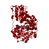
| |||||||||||||||||||||
| Unit cell |
| |||||||||||||||||||||
| Components on special symmetry positions |
|
-Components
| #1: Protein | Mass: 65351.508 Da / Num. of mol.: 1 Source method: isolated from a genetically manipulated source Source: (gene. exp.) Aerococcus viridans (bacteria) / Production host: | ||||||
|---|---|---|---|---|---|---|---|
| #2: Chemical | ChemComp-SO4 /   Mass: 96.063 Da / Num. of mol.: 4 / Source method: obtained synthetically / Formula: SO4 Mass: 96.063 Da / Num. of mol.: 4 / Source method: obtained synthetically / Formula: SO4#3: Chemical | ChemComp-FAD / |   Mass: 785.550 Da / Num. of mol.: 1 / Source method: obtained synthetically / Formula: C27H33N9O15P2 / Comment: FAD*YM Mass: 785.550 Da / Num. of mol.: 1 / Source method: obtained synthetically / Formula: C27H33N9O15P2 / Comment: FAD*YM#4: Water | ChemComp-HOH / |  Mass: 18.015 Da / Num. of mol.: 693 / Source method: isolated from a natural source / Formula: H2O Mass: 18.015 Da / Num. of mol.: 693 / Source method: isolated from a natural source / Formula: H2OSequence details | A SEQUENCE DATABASE REFERENCE FOR THIS PROTEIN DOES NOT CURRENTLY EXIST. | |
-Experimental details
-Experiment
| Experiment | Method: X-RAY DIFFRACTION / Number of used crystals: 1 |
|---|
- Sample preparation
| Crystal | Density Matthews: 2.42 Å3/Da / Density % sol: 49.28 % |
|---|---|
| Crystal grow | Temperature: 293 K / Method: vapor diffusion, hanging drop / pH: 7 Details: 2M ammonium sulfate in 20mM sodium phosphate buffer, pH 7.0., VAPOR DIFFUSION, HANGING DROP, temperature 293K |
-Data collection
| Diffraction | Mean temperature: 100 K |
|---|---|
| Diffraction source | Source: SYNCHROTRON / Site: Photon Factory  / Beamline: AR-NW12A / Wavelength: 1 Å / Beamline: AR-NW12A / Wavelength: 1 Å |
| Detector | Type: ADSC QUANTUM 210 / Detector: CCD / Date: Dec 17, 2003 |
| Radiation | Monochromator: SI(111) / Protocol: SINGLE WAVELENGTH / Monochromatic (M) / Laue (L): M / Scattering type: x-ray |
| Radiation wavelength | Wavelength: 1 Å / Relative weight: 1 |
| Reflection | Resolution: 1.58→50 Å / Num. obs: 84186 / % possible obs: 95.5 % / Observed criterion σ(F): 0 / Observed criterion σ(I): 0 / Redundancy: 5.7 % / Rmerge(I) obs: 0.082 |
| Reflection shell | Resolution: 1.58→1.64 Å / Redundancy: 2.5 % / Rmerge(I) obs: 0.379 / % possible all: 77.4 |
- Processing
| Software |
| |||||||||||||||||||||||||
|---|---|---|---|---|---|---|---|---|---|---|---|---|---|---|---|---|---|---|---|---|---|---|---|---|---|---|
| Refinement | Method to determine structure: MOLECULAR REPLACEMENT Starting model: 1POX Resolution: 1.6→10 Å / Cross valid method: THROUGHOUT / σ(F): 3 / Stereochemistry target values: Maximum Likelihood Details: THE AMINO ACID RESIDUES 471-491 HAVE HIGH B-FACTOR VALUES AND MOST OF THEIR ATOMS HAVE LOW OCCUPANCY. THIS REGION, WHICH IS ASSUMED TO HAVE HIGH FLEXIBILITY, IS ALSO FOUND IN OTHER THIAMIN ...Details: THE AMINO ACID RESIDUES 471-491 HAVE HIGH B-FACTOR VALUES AND MOST OF THEIR ATOMS HAVE LOW OCCUPANCY. THIS REGION, WHICH IS ASSUMED TO HAVE HIGH FLEXIBILITY, IS ALSO FOUND IN OTHER THIAMIN DIPHOSPHATE-DEPENDENT ENZYMES.
| |||||||||||||||||||||||||
| Displacement parameters |
| |||||||||||||||||||||||||
| Refinement step | Cycle: LAST / Resolution: 1.6→10 Å
|
