[English] 日本語
Yorodumi
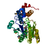
- PDB-4nqw: Structure of Mycobacterium tuberculosis extracytoplasmic function... -

+
Open data


ID or keywords:

Loading...

-
Basic information

Entry
Database: PDB / ID: 4nqw
Title
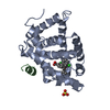
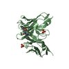
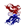
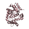
Structure of Mycobacterium tuberculosis extracytoplasmic function sigma factor SigK in complex with the cytosolic domain of its cognate anti-sigma factor RskA
Components
  • Anti-sigma-K factor RskA
  • ECF RNA polymerase sigma factor SigK
KeywordsDNA BINDING PROTEIN/PROTEIN BINDING / sigma factor / transcription initiation / DNA binding / Promoter DNA binding and transcription initiation / anti-sigma factor / DNA BINDING PROTEIN-PROTEIN BINDING complex
Function / homology
Function and homology information


sigma factor antagonist activity / cell wall / sigma factor activity / membrane => GO:0016020 / peptidoglycan-based cell wall / DNA-templated transcription initiation / regulation of translation / DNA-binding transcription factor activity / regulation of DNA-templated transcription / DNA binding / plasma membrane
Similarity search - Function
Anti-sigma K factor RskA / : / Anti-sigma-K factor rskA, C-terminal / Anti-sigma-K factor RskA, N-terminal domain / : / Anti-sigma factor, zinc-finger domain / Anti-sigma factor, zinc-finger domain superfamily / RNA polymerase sigma-70 like / RNA polymerase sigma factor, region 2, helix turn helix motif / Rna Polymerase Sigma Factor; Chain: A ...Anti-sigma K factor RskA / : / Anti-sigma-K factor rskA, C-terminal / Anti-sigma-K factor RskA, N-terminal domain / : / Anti-sigma factor, zinc-finger domain / Anti-sigma factor, zinc-finger domain superfamily / RNA polymerase sigma-70 like / RNA polymerase sigma factor, region 2, helix turn helix motif / Rna Polymerase Sigma Factor; Chain: A / RNA polymerase sigma-70 region 4 / Sigma-70, region 4 / RNA polymerase sigma-70 region 2 / RNA polymerase sigma-70 like domain / Sigma-70 region 2 / RNA polymerase sigma factor, region 2 / RNA polymerase sigma factor, region 3/4-like / Winged helix-like DNA-binding domain superfamily/Winged helix DNA-binding domain / Arc Repressor Mutant, subunit A / Winged helix-like DNA-binding domain superfamily / Orthogonal Bundle / Mainly Alpha
Similarity search - Domain/homology
: / Anti-sigma-K factor RskA / ECF RNA polymerase sigma factor SigK / ECF RNA polymerase sigma factor SigK / Anti-sigma-K factor RskA
Similarity search - Component
Biological speciesMycobacterium tuberculosis (bacteria)
MethodX-RAY DIFFRACTION / SAD / Resolution: 2.4 Å
AuthorsShukla, J.K. / Gopal, B.
CitationJournal: Acta Crystallogr.,Sect.D / Year: 2014
Title: Structural basis for the redox sensitivity of the Mycobacterium tuberculosis SigK-RskA sigma-anti-sigma complex
Authors: Shukla, J. / Gupta, R. / Thakur, K.G. / Gokhale, R. / Gopal, B.
History
DepositionNov 25, 2013Deposition site: RCSB / Processing site: PDBJ
SupersessionJan 22, 2014ID: 3VDO
Revision 1.0Jan 22, 2014Provider: repository / Type: Initial release
Revision 1.1Apr 16, 2014Group: Database references
Revision 1.2Nov 20, 2024Group: Data collection / Database references ...Data collection / Database references / Derived calculations / Structure summary
Category: chem_comp_atom / chem_comp_bond ...chem_comp_atom / chem_comp_bond / database_2 / pdbx_entry_details / pdbx_modification_feature / pdbx_struct_conn_angle / struct_conn / struct_ref_seq_dif / struct_site
Item: _database_2.pdbx_DOI / _database_2.pdbx_database_accession ..._database_2.pdbx_DOI / _database_2.pdbx_database_accession / _pdbx_struct_conn_angle.ptnr1_auth_asym_id / _pdbx_struct_conn_angle.ptnr1_auth_comp_id / _pdbx_struct_conn_angle.ptnr1_auth_seq_id / _pdbx_struct_conn_angle.ptnr1_label_asym_id / _pdbx_struct_conn_angle.ptnr1_label_atom_id / _pdbx_struct_conn_angle.ptnr1_label_comp_id / _pdbx_struct_conn_angle.ptnr1_label_seq_id / _pdbx_struct_conn_angle.ptnr2_auth_asym_id / _pdbx_struct_conn_angle.ptnr2_auth_seq_id / _pdbx_struct_conn_angle.ptnr2_label_asym_id / _pdbx_struct_conn_angle.ptnr3_auth_asym_id / _pdbx_struct_conn_angle.ptnr3_auth_comp_id / _pdbx_struct_conn_angle.ptnr3_auth_seq_id / _pdbx_struct_conn_angle.ptnr3_label_asym_id / _pdbx_struct_conn_angle.ptnr3_label_atom_id / _pdbx_struct_conn_angle.ptnr3_label_comp_id / _pdbx_struct_conn_angle.ptnr3_label_seq_id / _pdbx_struct_conn_angle.value / _struct_conn.pdbx_dist_value / _struct_conn.ptnr1_auth_asym_id / _struct_conn.ptnr1_auth_comp_id / _struct_conn.ptnr1_auth_seq_id / _struct_conn.ptnr1_label_asym_id / _struct_conn.ptnr1_label_atom_id / _struct_conn.ptnr1_label_comp_id / _struct_conn.ptnr1_label_seq_id / _struct_conn.ptnr2_auth_asym_id / _struct_conn.ptnr2_auth_comp_id / _struct_conn.ptnr2_auth_seq_id / _struct_conn.ptnr2_label_asym_id / _struct_conn.ptnr2_label_atom_id / _struct_conn.ptnr2_label_comp_id / _struct_conn.ptnr2_label_seq_id / _struct_ref_seq_dif.details / _struct_site.pdbx_auth_asym_id / _struct_site.pdbx_auth_comp_id / _struct_site.pdbx_auth_seq_id

-
Structure visualization

Structure viewerMolecule:
MolmilJmol/JSmol

Downloads & links

-
Assembly

Deposited unit
A: ECF RNA polymerase sigma factor SigK
B: Anti-sigma-K factor RskA
hetero molecules


Theoretical massNumber of molelcules
Total (without water)32,65112
Polymers31,5272
Non-polymers1,12410
Water1,60389
1


  • Idetical with deposited unit
  • defined by author&software
TypeNameSymmetry operationNumber
identity operation1_555x,y,z1
Buried area5160 Å2
ΔGint-85 kcal/mol
Surface area11300 Å2
MethodPISA
Unit cell
Length a, b, c (Å)57.390, 57.390, 150.870
Angle α, β, γ (deg.)90.00, 90.00, 90.00
Int Tables number92
Space group name H-MP41212

-
Components

#1: Protein ECF RNA polymerase sigma factor SigK / ECF sigma factor SigK / Alternative RNA polymerase sigma factor SigK / RNA polymerase sigma-K ...ECF sigma factor SigK / Alternative RNA polymerase sigma factor SigK / RNA polymerase sigma-K factor / Sigma-K factor


Mass: 22971.732 Da / Num. of mol.: 1
Source method: isolated from a genetically manipulated source
Source: (gene. exp.) Mycobacterium tuberculosis (bacteria) / Strain: H37Rv / Gene: Rv0445c, sigK / Plasmid: pETDUET-1 / Production host: Escherichia coli (E. coli) / Strain (production host): Rosetta (DE3) / References: UniProt: O53730, UniProt: P9WGH7*PLUS
#2: Protein Anti-sigma-K factor RskA / Regulator of SigK / Sigma-K anti-sigma factor RskA


Mass: 8555.508 Da / Num. of mol.: 1 / Fragment: UNP residues 1-80
Source method: isolated from a genetically manipulated source
Source: (gene. exp.) Mycobacterium tuberculosis (bacteria) / Strain: H37Rv / Gene: rskA, Rv0444c / Plasmid: pETDUET-1 / Production host: Escherichia coli (E. coli) / Strain (production host): Rosetta (DE3) / References: UniProt: O53729, UniProt: P9WGX4*PLUS
#3: Chemical
ChemComp-CD / CADMIUM ION


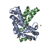
Mass: 112.411 Da / Num. of mol.: 10 / Source method: obtained synthetically / Formula: Cd
#4: Water ChemComp-HOH / water


Mass: 18.015 Da / Num. of mol.: 89 / Source method: isolated from a natural source / Formula: H2O
Has protein modificationY

-
Experimental details

-
Experiment

ExperimentMethod: X-RAY DIFFRACTION / Number of used crystals: 1

-
Sample preparation

CrystalDensity Matthews: 1.97 Å3/Da / Density % sol: 37.57 %
Crystal growTemperature: 295 K / Method: microbatch underoil / pH: 7.5
Details: 0.1M HEPES, 1M sodium acetate trihydrate, 0.05M cadmium sulphate hydrate, 12%(v/v) 2,5 hexanediol, pH 7.5, Microbatch underoil, temperature 295K

-
Data collection

DiffractionMean temperature: 100 K
Diffraction sourceSource: ROTATING ANODE / Type: RIGAKU MICROMAX-007 HF / Wavelength: 1.5418 Å
DetectorType: MAR scanner 345 mm plate / Detector: IMAGE PLATE / Date: Jan 28, 2010
RadiationMonochromator: VariMaxHF / Protocol: SINGLE WAVELENGTH / Monochromatic (M) / Laue (L): M / Scattering type: x-ray
Radiation wavelengthWavelength: 1.5418 Å / Relative weight: 1
ReflectionResolution: 2.4→28.695 Å / Num. all: 9753 / Num. obs: 9747 / Redundancy: 14.8 % / Rmerge(I) obs: 0.093 / Net I/σ(I): 20.1
Reflection shellResolution: 2.4→2.53 Å / Rmerge(I) obs: 0.339 / Mean I/σ(I) obs: 8.1 / Num. unique all: 1480 / % possible all: 100

-
Processing

Software
NameVersionClassification
MAR345dtbdata collection
SOLVEphasing
REFMAC5.7.0029refinement
MOSFLMdata reduction
SCALAdata scaling
RefinementMethod to determine structure: SAD / Resolution: 2.4→28.69 Å / Cor.coef. Fo:Fc: 0.927 / Cor.coef. Fo:Fc free: 0.909 / SU B: 20.303 / SU ML: 0.208 / Cross valid method: THROUGHOUT / ESU R Free: 0.317 / Stereochemistry target values: MAXIMUM LIKELIHOOD / Details: HYDROGENS HAVE BEEN ADDED IN THE RIDING POSITIONS
RfactorNum. reflection% reflectionSelection details
Rfree0.25986 916 9.9 %RANDOM
Rwork0.20463 ---
obs0.21018 8305 87.82 %-
Solvent computationIon probe radii: 0.8 Å / Shrinkage radii: 0.8 Å / VDW probe radii: 1.2 Å / Solvent model: MASK
Displacement parametersBiso mean: 32.018 Å2
Baniso -1Baniso -2Baniso -3
1-0.05 Å20 Å20 Å2
2--0.05 Å2-0 Å2
3----0.1 Å2
Refinement stepCycle: LAST / Resolution: 2.4→28.69 Å
ProteinNucleic acidLigandSolventTotal
Num. atoms1740 0 10 89 1839
Refine LS restraints
Refine-IDTypeDev idealDev ideal targetNumber
X-RAY DIFFRACTIONr_bond_refined_d0.0050.0191765
X-RAY DIFFRACTIONr_bond_other_d0.0010.021700
X-RAY DIFFRACTIONr_angle_refined_deg0.8421.9562387
X-RAY DIFFRACTIONr_angle_other_deg0.7273.0013864
X-RAY DIFFRACTIONr_dihedral_angle_1_deg4.0525218
X-RAY DIFFRACTIONr_dihedral_angle_2_deg29.25321.72487
X-RAY DIFFRACTIONr_dihedral_angle_3_deg16.17215298
X-RAY DIFFRACTIONr_dihedral_angle_4_deg11.9571526
X-RAY DIFFRACTIONr_chiral_restr0.0460.2276
X-RAY DIFFRACTIONr_gen_planes_refined0.0030.021992
X-RAY DIFFRACTIONr_gen_planes_other0.0010.02424
X-RAY DIFFRACTIONr_rigid_bond_restr1.60233465
X-RAY DIFFRACTIONr_sphericity_free33.799536
X-RAY DIFFRACTIONr_sphericity_bonded12.08853504
LS refinement shellResolution: 2.4→2.462 Å / Total num. of bins used: 20
RfactorNum. reflection% reflection
Rfree0.318 71 -
Rwork0.218 637 -
obs--94.65 %
Refinement TLS params.

Method: refined / Refine-ID: X-RAY DIFFRACTION

IDL112)L122)L132)L222)L232)L332)S11 (Å °)S12 (Å °)S13 (Å °)S21 (Å °)S22 (Å °)S23 (Å °)S31 (Å °)S32 (Å °)S33 (Å °)T112)T122)T132)T222)T232)T332)Origin x (Å)Origin y (Å)Origin z (Å)
10.43450.1811-0.33940.9101-1.07791.31660.04660.0443-0.00070.0680.01450.0494-0.0891-0.0292-0.06110.05070.0019-0.00820.0295-0.01390.014315.65056.456846.9331
20.17190.2205-0.18721.4819-1.10920.85250.0086-0.0370.0062-0.02660.05150.1057-0.0074-0.0344-0.06010.0431-0.0021-0.01580.0822-0.00660.02788.91892.480246.7229
Refinement TLS group
IDRefine-IDRefine TLS-IDAuth asym-IDAuth seq-ID
1X-RAY DIFFRACTION1A10 - 185
2X-RAY DIFFRACTION2B7 - 79

+
About Yorodumi

-
News

-
Feb 9, 2022. New format data for meta-information of EMDB entries

New format data for meta-information of EMDB entries

  • Version 3 of the EMDB header file is now the official format.
  • The previous official version 1.9 will be removed from the archive.

Related info.:EMDB header

External links:wwPDB to switch to version 3 of the EMDB data model

-
Aug 12, 2020. Covid-19 info

Covid-19 info

URL: https://pdbj.org/emnavi/covid19.php

New page: Covid-19 featured information page in EM Navigator.

Related info.:Covid-19 info / Mar 5, 2020. Novel coronavirus structure data

+
Mar 5, 2020. Novel coronavirus structure data

Novel coronavirus structure data

Related info.:Yorodumi Speices / Aug 12, 2020. Covid-19 info

External links:COVID-19 featured content - PDBj / Molecule of the Month (242):Coronavirus Proteases

+
Jan 31, 2019. EMDB accession codes are about to change! (news from PDBe EMDB page)

EMDB accession codes are about to change! (news from PDBe EMDB page)

  • The allocation of 4 digits for EMDB accession codes will soon come to an end. Whilst these codes will remain in use, new EMDB accession codes will include an additional digit and will expand incrementally as the available range of codes is exhausted. The current 4-digit format prefixed with “EMD-” (i.e. EMD-XXXX) will advance to a 5-digit format (i.e. EMD-XXXXX), and so on. It is currently estimated that the 4-digit codes will be depleted around Spring 2019, at which point the 5-digit format will come into force.
  • The EM Navigator/Yorodumi systems omit the EMD- prefix.

Related info.:Q: What is EMD? / ID/Accession-code notation in Yorodumi/EM Navigator

External links:EMDB Accession Codes are Changing Soon! / Contact to PDBj

+
Jul 12, 2017. Major update of PDB

Major update of PDB

  • wwPDB released updated PDB data conforming to the new PDBx/mmCIF dictionary.
  • This is a major update changing the version number from 4 to 5, and with Remediation, in which all the entries are updated.
  • In this update, many items about electron microscopy experimental information are reorganized (e.g. em_software).
  • Now, EM Navigator and Yorodumi are based on the updated data.

External links:wwPDB Remediation / Enriched Model Files Conforming to OneDep Data Standards Now Available in the PDB FTP Archive

-
Yorodumi

Thousand views of thousand structures

  • Yorodumi is a browser for structure data from EMDB, PDB, SASBDB, etc.
  • This page is also the successor to EM Navigator detail page, and also detail information page/front-end page for Omokage search.
  • The word "yorodu" (or yorozu) is an old Japanese word meaning "ten thousand". "mi" (miru) is to see.

Related info.:EMDB / PDB / SASBDB / Comparison of 3 databanks / Yorodumi Search / Aug 31, 2016. New EM Navigator & Yorodumi / Yorodumi Papers / Jmol/JSmol / Function and homology information / Changes in new EM Navigator and Yorodumi

Read more