 Movie
Movie Controller

[English] 日本語
 Yorodumi
- PDB-4nmi: Crystal Structure of the Apo ectoine hydroxylase ECTD from Saliba... -
+ Open data

- Basic information
| Entry | Database: PDB / ID: 4nmi | ||||||
|---|---|---|---|---|---|---|---|
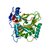
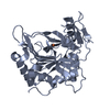
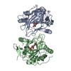
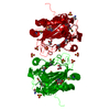
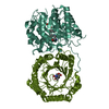
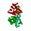
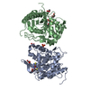
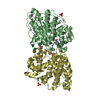
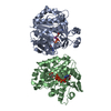
| Title | Crystal Structure of the Apo ectoine hydroxylase ECTD from Salibacillus salexigens | ||||||
 Components Components | EctD | ||||||
 Keywords Keywords | OXIDOREDUCTASE / jelly-roll or cupin fold / metal ion binding / Iron | ||||||
| Function / homology |  Function and homology information Function and homology informationectoine hydroxylase / ectoine catabolic process / 2-oxoglutarate-dependent dioxygenase activity / iron ion binding Similarity search - Function | ||||||
| Biological species |  Virgibacillus salexigens (bacteria) Virgibacillus salexigens (bacteria) | ||||||
| Method |  X-RAY DIFFRACTION / SYNCHROTRON / MOLECULAR REPLACEMENT / Resolution: 1.78 Å X-RAY DIFFRACTION / SYNCHROTRON / MOLECULAR REPLACEMENT / Resolution: 1.78 Å | ||||||
 Authors Authors | Widderich, N. / Hoeppner, A. / Smits, S.H. / Bremer, E. | ||||||
 Citation Citation | Journal: Plos One / Year: 2014 Title: Biochemical properties of ectoine hydroxylases from extremophiles and their wider taxonomic distribution among microorganisms. Authors: Widderich, N. / Hoppner, A. / Pittelkow, M. / Heider, J. / Smits, S.H. / Bremer, E. | ||||||
| History |
|
- Structure visualization
| Structure viewer | Molecule: MolmilJmol/JSmol |
|---|
- Downloads & links
-Download
| PDBx/mmCIF format | 4nmi.cif.gz | 77.3 KB | Display | PDBx/mmCIF format |
|---|---|---|---|---|
| PDB format | pdb4nmi.ent.gz | 57.8 KB | Display | PDB format |
| PDBx/mmJSON format | 4nmi.json.gz | Tree view | PDBx/mmJSON format | |
| Others |  Other downloads Other downloads |
-Validation report
| Arichive directory | https://data.pdbj.org/pub/pdb/validation_reports/nm/4nmiftp://data.pdbj.org/pub/pdb/validation_reports/nm/4nmi | HTTPS FTP |
|---|
-Related structure data
| Related structure data | |
|---|---|
| Similar structure data |


-Links
 PDBj
PDBj- Assembly
| Deposited unit | 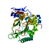
| ||||||||
|---|---|---|---|---|---|---|---|---|---|
| 1 | 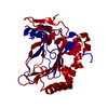
| ||||||||
| Unit cell |
| ||||||||
| Components on special symmetry positions |
|
-Components
| #1: Protein | Mass: 35651.836 Da / Num. of mol.: 1 / Fragment: ectoine hydroxylase / Source method: isolated from a natural source / Source: (natural) Virgibacillus salexigens (bacteria)References: UniProt: Q2TDY4, Oxidoreductases; Acting on paired donors, with incorporation or reduction of molecular oxygen; With 2-oxoglutarate as one donor, and incorporation of one atom of oxygen into each donor |
|---|---|
| #2: Water | ChemComp-HOH /  Mass: 18.015 Da / Num. of mol.: 349 / Source method: isolated from a natural source / Formula: H2O Mass: 18.015 Da / Num. of mol.: 349 / Source method: isolated from a natural source / Formula: H2O |
-Experimental details
-Experiment
| Experiment | Method: X-RAY DIFFRACTION / Number of used crystals: 1 |
|---|
- Sample preparation
| Crystal | Density Matthews: 3.42 Å3/Da / Density % sol: 64.05 % |
|---|---|
| Crystal grow | Temperature: 298 K / Method: vapor diffusion, hanging drop / pH: 5 Details: 100 mM MES pH 5.0 and 1.2 M ammonium sulfate , VAPOR DIFFUSION, HANGING DROP, temperature 298K |
-Data collection
| Diffraction | Mean temperature: 200 K |
|---|---|
| Diffraction source | Source: SYNCHROTRON / Site: ESRF  / Beamline: ID23-1 / Wavelength: 0.92 Å / Beamline: ID23-1 / Wavelength: 0.92 Å |
| Detector | Type: MAR CCD 165 mm / Detector: CCD / Date: Aug 14, 2013 |
| Radiation | Monochromator: Ni FILTER / Protocol: SINGLE WAVELENGTH / Monochromatic (M) / Laue (L): M / Scattering type: x-ray |
| Radiation wavelength | Wavelength: 0.92 Å / Relative weight: 1 |
| Reflection | Resolution: 1.7→20 Å / Num. all: 40025 / Num. obs: 37155 / % possible obs: 92.8 % / Observed criterion σ(F): 2 / Observed criterion σ(I): 2 |
| Reflection shell | Resolution: 1.78→1.826 Å / % possible all: 90.9 |
- Processing
| Software |
| ||||||||||||||||||||||||||||||||||||||||||||||||||||||||||||
|---|---|---|---|---|---|---|---|---|---|---|---|---|---|---|---|---|---|---|---|---|---|---|---|---|---|---|---|---|---|---|---|---|---|---|---|---|---|---|---|---|---|---|---|---|---|---|---|---|---|---|---|---|---|---|---|---|---|---|---|---|---|
| Refinement | Method to determine structure: MOLECULAR REPLACEMENT / Resolution: 1.78→20 Å / Cor.coef. Fo:Fc: 0.963 / Cor.coef. Fo:Fc free: 0.952 / SU B: 1.798 / SU ML: 0.056 / Cross valid method: THROUGHOUT / σ(F): 2 / ESU R: 0.084 / ESU R Free: 0.086 / Stereochemistry target values: MAXIMUM LIKELIHOOD / Details: HYDROGENS HAVE BEEN ADDED IN THE RIDING POSITIONS
| ||||||||||||||||||||||||||||||||||||||||||||||||||||||||||||
| Solvent computation | Ion probe radii: 0.8 Å / Shrinkage radii: 0.8 Å / VDW probe radii: 1.2 Å / Solvent model: MASK | ||||||||||||||||||||||||||||||||||||||||||||||||||||||||||||
| Displacement parameters | Biso mean: 25.955 Å2
| ||||||||||||||||||||||||||||||||||||||||||||||||||||||||||||
| Refine analyze |
| ||||||||||||||||||||||||||||||||||||||||||||||||||||||||||||
| Refinement step | Cycle: LAST / Resolution: 1.78→20 Å
| ||||||||||||||||||||||||||||||||||||||||||||||||||||||||||||
| Refine LS restraints |
| ||||||||||||||||||||||||||||||||||||||||||||||||||||||||||||
| LS refinement shell | Resolution: 1.78→1.826 Å / Total num. of bins used: 20
|
