 Movie
Movie Controller

[English] 日本語
 Yorodumi
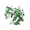
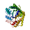
- PDB-3emr: Crystal Structure Analysis of the ectoine hydroxylase ECTD from S... -
+ Open data

- Basic information
| Entry | Database: PDB / ID: 3emr | ||||||
|---|---|---|---|---|---|---|---|
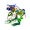
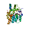
| Title | Crystal Structure Analysis of the ectoine hydroxylase ECTD from Salibacillus salexigens | ||||||
 Components Components | EctD | ||||||
 Keywords Keywords | OXIDOREDUCTASE / double stranded beta helix | ||||||
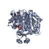
| Function / homology |  Function and homology information Function and homology informationectoine hydroxylase / ectoine catabolic process / 2-oxoglutarate-dependent dioxygenase activity / iron ion binding Similarity search - Function | ||||||
| Biological species |  Virgibacillus salexigens (bacteria) Virgibacillus salexigens (bacteria) | ||||||
| Method |  X-RAY DIFFRACTION / SYNCHROTRON / MAD / Resolution: 1.85 Å X-RAY DIFFRACTION / SYNCHROTRON / MAD / Resolution: 1.85 Å | ||||||
 Authors Authors | Reuter, K. / Heine, A. | ||||||
 Citation Citation | Journal: Plos One / Year: 2010 Title: Synthesis of 5-hydroxyectoine from ectoine: crystal structure of the non-heme iron(II) and 2-oxoglutarate-dependent dioxygenase EctD Authors: Reuter, K. / Pittelkow, M. / Bursy, J. / Heine, A. / Craan, T. / Bremer, E. | ||||||
| History |
|
- Structure visualization
| Structure viewer | Molecule: MolmilJmol/JSmol |
|---|
- Downloads & links
-Download
| PDBx/mmCIF format | 3emr.cif.gz | 77.3 KB | Display | PDBx/mmCIF format |
|---|---|---|---|---|
| PDB format | pdb3emr.ent.gz | 56.4 KB | Display | PDB format |
| PDBx/mmJSON format | 3emr.json.gz | Tree view | PDBx/mmJSON format | |
| Others |  Other downloads Other downloads |
-Validation report
| Arichive directory | https://data.pdbj.org/pub/pdb/validation_reports/em/3emrftp://data.pdbj.org/pub/pdb/validation_reports/em/3emr | HTTPS FTP |
|---|
-Related structure data
| Similar structure data |
|---|


-Links
 PDBj
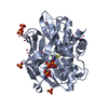
PDBj- Assembly
| Deposited unit | 
| |||||||||
|---|---|---|---|---|---|---|---|---|---|---|
| 1 |
| |||||||||
| 2 | 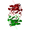
| |||||||||
| Unit cell |
| |||||||||
| Components on special symmetry positions |
| |||||||||
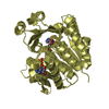
| Details | ECTD HAS CLEARLY BEEN SHOWN BY SIZE EXCLUSION CHROMATOGRAPHY TO BE A MONOMER IN SOLUTION (BURSY ET AL. 2007, J. BIOL. CHEM. 282, PP31147-31155), AGAINST PISA'S SUGGESTION, A HOMODIMER. |
-Components
| #1: Protein | Mass: 35980.098 Da / Num. of mol.: 1 Source method: isolated from a genetically manipulated source Details: cell / Source: (gene. exp.) Virgibacillus salexigens (bacteria) / Strain: DSM-11483 / Gene: ectD / Plasmid: pASK-IBA3 / Production host: References: UniProt: Q2TDY4, Oxidoreductases; Acting on paired donors, with incorporation or reduction of molecular oxygen; With 2-oxoglutarate as one donor, and incorporation of one atom of oxygen into each donor | ||||||
|---|---|---|---|---|---|---|---|
| #2: Chemical | ChemComp-FE /   Mass: 55.845 Da / Num. of mol.: 1 / Source method: obtained synthetically / Formula: Fe Mass: 55.845 Da / Num. of mol.: 1 / Source method: obtained synthetically / Formula: Fe | ||||||
| #3: Chemical | ChemComp-SO4 /   Mass: 96.063 Da / Num. of mol.: 4 / Source method: obtained synthetically / Formula: SO4 Mass: 96.063 Da / Num. of mol.: 4 / Source method: obtained synthetically / Formula: SO4#4: Chemical |   Mass: 92.094 Da / Num. of mol.: 2 / Source method: obtained synthetically / Formula: C3H8O3 Mass: 92.094 Da / Num. of mol.: 2 / Source method: obtained synthetically / Formula: C3H8O3#5: Water | ChemComp-HOH / |  Mass: 18.015 Da / Num. of mol.: 197 / Source method: isolated from a natural source / Formula: H2O Mass: 18.015 Da / Num. of mol.: 197 / Source method: isolated from a natural source / Formula: H2OHas protein modification | Y | |
-Experimental details
-Experiment
| Experiment | Method: X-RAY DIFFRACTION / Number of used crystals: 2 |
|---|
- Sample preparation
| Crystal | Density Matthews: 3.7 Å3/Da / Density % sol: 66.9 % |
|---|---|
| Crystal grow | Temperature: 291 K / Method: vapor diffusion, hanging drop / pH: 5 Details: 1.0M ammonium sulfate, 0.1M sodium fluoride, 2mM TCEP, 0.1M sodium acetate, pH 5.0, VAPOR DIFFUSION, HANGING DROP, temperature 291K |
-Data collection
| Diffraction |
| ||||||||||||||||||
|---|---|---|---|---|---|---|---|---|---|---|---|---|---|---|---|---|---|---|---|
| Diffraction source |
| ||||||||||||||||||
| Detector |
| ||||||||||||||||||
| Radiation |
| ||||||||||||||||||
| Radiation wavelength |
| ||||||||||||||||||
| Reflection | Resolution: 1.85→50 Å / Num. all: 43040 / Num. obs: 43040 / % possible obs: 99.9 % / Redundancy: 14.6 % / Rsym value: 0.067 / Net I/σ(I): 34.1 | ||||||||||||||||||
| Reflection shell | Resolution: 1.85→1.88 Å / Redundancy: 13.9 % / Mean I/σ(I) obs: 5.9 / Num. unique all: 2095 / Rsym value: 0.437 / % possible all: 100 |

- Processing
| Software |
| |||||||||||||||||||||||||||||||||
|---|---|---|---|---|---|---|---|---|---|---|---|---|---|---|---|---|---|---|---|---|---|---|---|---|---|---|---|---|---|---|---|---|---|---|
| Refinement | Method to determine structure: MAD / Resolution: 1.85→10 Å / Num. parameters: 9863 / Num. restraintsaints: 9313 / Cross valid method: FREE R / σ(F): 0 / σ(I): 0 / Stereochemistry target values: ENGH AND HUBER Details: ANISOTROPIC SCALING APPLIED BY THE METHOD OF PARKIN, MOEZZI & HOPE, J.APPL.CRYST.28(1995)53-56 ANISOTROPIC REFINEMENT REDUCED FREE R (NO CUTOFF) BY ?
| |||||||||||||||||||||||||||||||||
| Refine analyze | Num. disordered residues: 6 / Occupancy sum hydrogen: 2071 / Occupancy sum non hydrogen: 2421.1 | |||||||||||||||||||||||||||||||||
| Refinement step | Cycle: LAST / Resolution: 1.85→10 Å
| |||||||||||||||||||||||||||||||||
| Refine LS restraints |
|
