 Movie
Movie Controller

[English] 日本語
 Yorodumi
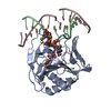
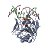
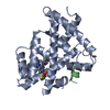
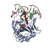
- PDB-4nii: Crystal structure of AlkB D135I mutant protein with cofactors bou... -
+ Open data

- Basic information
| Entry | Database: PDB / ID: 4nii | ||||||
|---|---|---|---|---|---|---|---|
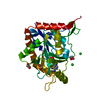
| Title | Crystal structure of AlkB D135I mutant protein with cofactors bound to dsDNA containing m6A/A | ||||||
 Components Components |
| ||||||
 Keywords Keywords | OXIDOREDUCTASE/DNA / DNA/RNA direct repair / jelly-roll fold / DNA/RNA demethylation repair / Fe(II) / 2-KG / OXIDOREDUCTASE-DNA complex | ||||||
| Function / homology |  Function and homology information Function and homology informationresponse to methyl methanesulfonate / oxidative RNA demethylation / DNA oxidative demethylase / broad specificity oxidative DNA demethylase activity / oxidative RNA demethylase activity / RNA repair / oxidative demethylation / DNA alkylation repair / dioxygenase activity / ferrous iron binding ...response to methyl methanesulfonate / oxidative RNA demethylation / DNA oxidative demethylase / broad specificity oxidative DNA demethylase activity / oxidative RNA demethylase activity / RNA repair / oxidative demethylation / DNA alkylation repair / dioxygenase activity / ferrous iron binding / DNA repair / cytosol / cytoplasm Similarity search - Function | ||||||
| Biological species |  | ||||||
| Method |  X-RAY DIFFRACTION / SYNCHROTRON / MOLECULAR REPLACEMENT / Resolution: 1.622 Å X-RAY DIFFRACTION / SYNCHROTRON / MOLECULAR REPLACEMENT / Resolution: 1.622 Å | ||||||
 Authors Authors | Yi, C.Q. / Zhu, C.X. | ||||||
 Citation Citation | Journal: Angew.Chem.Int.Ed.Engl. / Year: 2014 Title: Switching Demethylation Activities between AlkB Family RNA/DNA Demethylases through Exchange of Active-Site Residues. Authors: Zhu, C. / Yi, C. | ||||||
| History |
|
- Structure visualization
| Structure viewer | Molecule: MolmilJmol/JSmol |
|---|
- Downloads & links
-Download
| PDBx/mmCIF format | 4nii.cif.gz | 128.4 KB | Display | PDBx/mmCIF format |
|---|---|---|---|---|
| PDB format | pdb4nii.ent.gz | 95.7 KB | Display | PDB format |
| PDBx/mmJSON format | 4nii.json.gz | Tree view | PDBx/mmJSON format | |
| Others |  Other downloads Other downloads |
-Validation report
| Arichive directory | https://data.pdbj.org/pub/pdb/validation_reports/ni/4niiftp://data.pdbj.org/pub/pdb/validation_reports/ni/4nii | HTTPS FTP |
|---|
-Related structure data
| Related structure data | 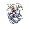 4nidC 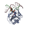 4nigC 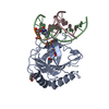 4nihC 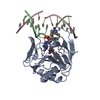 3bieS C: citing same article ( S: Starting model for refinement |
|---|---|
| Similar structure data |


-Links
 PDBj
PDBj


- Assembly
| Deposited unit | 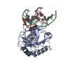
| ||||||||
|---|---|---|---|---|---|---|---|---|---|
| 1 |
| ||||||||
| Unit cell |
|
-Components
-Protein , 1 types, 1 molecules A
| #1: Protein | Mass: 22828.174 Da / Num. of mol.: 1 / Fragment: UNP residues 12-215 / Mutation: S129C/D135I Source method: isolated from a genetically manipulated source Source: (gene. exp.) |
|---|
-DNA chain , 2 types, 2 molecules BC
| #2: DNA chain | Mass: 4073.779 Da / Num. of mol.: 1 / Source method: obtained synthetically |
|---|---|
| #3: DNA chain | Mass: 3950.598 Da / Num. of mol.: 1 / Source method: obtained synthetically |
-Non-polymers , 3 types, 330 molecules 


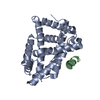
| #4: Chemical | ChemComp-MN /  Mass: 54.938 Da / Num. of mol.: 1 / Source method: obtained synthetically / Formula: Mn Mass: 54.938 Da / Num. of mol.: 1 / Source method: obtained synthetically / Formula: Mn |
|---|---|
| #5: Chemical | ChemComp-AKG /  Mass: 146.098 Da / Num. of mol.: 1 / Source method: obtained synthetically / Formula: C5H6O5 Mass: 146.098 Da / Num. of mol.: 1 / Source method: obtained synthetically / Formula: C5H6O5 |
| #6: Water | ChemComp-HOH / Mass: 18.015 Da / Num. of mol.: 328 / Source method: isolated from a natural source / Formula: H2O |
-Details
| Has protein modification | Y |
|---|
-Experimental details
-Experiment
| Experiment | Method: X-RAY DIFFRACTION / Number of used crystals: 1 |
|---|
- Sample preparation
| Crystal | Density Matthews: 2.54 Å3/Da / Density % sol: 51.53 % |
|---|---|
| Crystal grow | Temperature: 277 K / Method: vapor diffusion, hanging drop / pH: 6.5 Details: 20% PEG4000, 0.1 M sodium chloride, 0.05 M magnesium chloride, 0.1 M cacodylate, pH 6.5, VAPOR DIFFUSION, HANGING DROP, temperature 277K |
-Data collection
| Diffraction | Mean temperature: 200 K |
|---|---|
| Diffraction source | Source: SYNCHROTRON / Site: SSRF  / Beamline: BL17U / Wavelength: 0.97 Å / Beamline: BL17U / Wavelength: 0.97 Å |
| Detector | Type: ADSC QUANTUM 315r / Detector: CCD / Date: May 31, 2013 |
| Radiation | Monochromator: double crystal Si(111) / Protocol: SINGLE WAVELENGTH / Monochromatic (M) / Laue (L): M / Scattering type: x-ray |
| Radiation wavelength | Wavelength: 0.97 Å / Relative weight: 1 |
| Reflection | Resolution: 1.622→50 Å / Num. obs: 36703 / Observed criterion σ(F): 2.2 / Observed criterion σ(I): 2.2 |
| Reflection shell | Resolution: 1.622→1.69 Å / % possible all: 100 |
- Processing
| Software |
| ||||||||||||||||||||||||||||||||||||||||||||||||||||||||||||||||||||||||||||||||||||||||||||||||||||||||||||||||||||||||||||||||||||||||||||||||||||||||||||||||||||||||||||||||||||||
|---|---|---|---|---|---|---|---|---|---|---|---|---|---|---|---|---|---|---|---|---|---|---|---|---|---|---|---|---|---|---|---|---|---|---|---|---|---|---|---|---|---|---|---|---|---|---|---|---|---|---|---|---|---|---|---|---|---|---|---|---|---|---|---|---|---|---|---|---|---|---|---|---|---|---|---|---|---|---|---|---|---|---|---|---|---|---|---|---|---|---|---|---|---|---|---|---|---|---|---|---|---|---|---|---|---|---|---|---|---|---|---|---|---|---|---|---|---|---|---|---|---|---|---|---|---|---|---|---|---|---|---|---|---|---|---|---|---|---|---|---|---|---|---|---|---|---|---|---|---|---|---|---|---|---|---|---|---|---|---|---|---|---|---|---|---|---|---|---|---|---|---|---|---|---|---|---|---|---|---|---|---|---|---|
| Refinement | Method to determine structure: MOLECULAR REPLACEMENT Starting model: PDB ENTRY 3BIE Resolution: 1.622→37.06 Å / Cor.coef. Fo:Fc: 0.966 / Cor.coef. Fo:Fc free: 0.948 / SU B: 4.072 / SU ML: 0.07 / Cross valid method: THROUGHOUT / ESU R: 0.088 / ESU R Free: 0.09 / Stereochemistry target values: MAXIMUM LIKELIHOOD / Details: HYDROGENS HAVE BEEN ADDED IN THE RIDING POSITIONS
| ||||||||||||||||||||||||||||||||||||||||||||||||||||||||||||||||||||||||||||||||||||||||||||||||||||||||||||||||||||||||||||||||||||||||||||||||||||||||||||||||||||||||||||||||||||||
| Solvent computation | Ion probe radii: 0.8 Å / Shrinkage radii: 0.8 Å / VDW probe radii: 1.2 Å / Solvent model: MASK | ||||||||||||||||||||||||||||||||||||||||||||||||||||||||||||||||||||||||||||||||||||||||||||||||||||||||||||||||||||||||||||||||||||||||||||||||||||||||||||||||||||||||||||||||||||||
| Displacement parameters | Biso mean: 20.409 Å2
| ||||||||||||||||||||||||||||||||||||||||||||||||||||||||||||||||||||||||||||||||||||||||||||||||||||||||||||||||||||||||||||||||||||||||||||||||||||||||||||||||||||||||||||||||||||||
| Refinement step | Cycle: LAST / Resolution: 1.622→37.06 Å
| ||||||||||||||||||||||||||||||||||||||||||||||||||||||||||||||||||||||||||||||||||||||||||||||||||||||||||||||||||||||||||||||||||||||||||||||||||||||||||||||||||||||||||||||||||||||
| Refine LS restraints |
|