 Movie
Movie Controller

[English] 日本語
 Yorodumi
- PDB-4nd2: Crystal structure of the lactate dehydrogenase from cryptosporidi... -
+ Open data

- Basic information
| Entry | Database: PDB / ID: 4nd2 | ||||||||||||
|---|---|---|---|---|---|---|---|---|---|---|---|---|---|
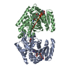
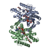
| Title | Crystal structure of the lactate dehydrogenase from cryptosporidium parvum complexed with substrate (pyruvic acid) and cofactor analog (3-acetylpyridine adenine dinucleotide) | ||||||||||||
 Components Components | Lactate dehydrogenase, adjacent gene encodes predicted malate dehydrogenase | ||||||||||||
 Keywords Keywords | OXIDOREDUCTASE / Rossmann Fold / Dehydrogenase / NAD Binding | ||||||||||||
| Function / homology |  Function and homology information Function and homology informationL-lactate dehydrogenase (NAD+) activity / lactate metabolic process / nucleotide binding Similarity search - Function | ||||||||||||
| Biological species |   Cryptosporidium parvum (eukaryote) Cryptosporidium parvum (eukaryote) | ||||||||||||
| Method |  X-RAY DIFFRACTION / SYNCHROTRON / MOLECULAR REPLACEMENT / Resolution: 2 Å X-RAY DIFFRACTION / SYNCHROTRON / MOLECULAR REPLACEMENT / Resolution: 2 Å | ||||||||||||
 Authors Authors | Chattopadhyay, D. / Cook, W.J. | ||||||||||||
 Citation Citation | Journal: Int.J.Biol.Macromol. / Year: 2014 Title: Biochemical and structural characterization of Cryptosporidium parvum Lactate dehydrogenase. Authors: Cook, W.J. / Senkovich, O. / Hernandez, A. / Speed, H. / Chattopadhyay, D. | ||||||||||||
| History |
|
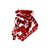
- Structure visualization
| Structure viewer | Molecule: MolmilJmol/JSmol |
|---|
- Downloads & links
-Download
| PDBx/mmCIF format | 4nd2.cif.gz | 142.1 KB | Display | PDBx/mmCIF format |
|---|---|---|---|---|
| PDB format | pdb4nd2.ent.gz | 110.7 KB | Display | PDB format |
| PDBx/mmJSON format | 4nd2.json.gz | Tree view | PDBx/mmJSON format | |
| Others |  Other downloads Other downloads |
-Validation report
| Arichive directory | https://data.pdbj.org/pub/pdb/validation_reports/nd/4nd2ftp://data.pdbj.org/pub/pdb/validation_reports/nd/4nd2 | HTTPS FTP |
|---|
-Related structure data
| Related structure data |  4nd1C  4nd3C  4nd4C  4nd5C  1t2dS C: citing same article ( S: Starting model for refinement |
|---|---|
| Similar structure data |


-Links
 PDBj
PDBj


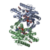
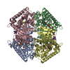
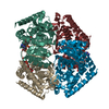
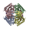
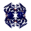
- Assembly
| Deposited unit | 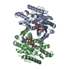
| ||||||||
|---|---|---|---|---|---|---|---|---|---|
| 1 | 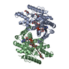
| ||||||||
| Unit cell |
|
-Components
| #1: Protein | Mass: 33884.855 Da / Num. of mol.: 2 / Fragment: unp residues 17-337 Source method: isolated from a genetically manipulated source Source: (gene. exp.) Cryptosporidium parvum (eukaryote) / Strain: Iowa II / Gene: cgd7_480, LDH1 / Plasmid: PET21A / Production host:  #2: Chemical |   Mass: 662.437 Da / Num. of mol.: 2 / Source method: obtained synthetically / Formula: C22H28N6O14P2 Mass: 662.437 Da / Num. of mol.: 2 / Source method: obtained synthetically / Formula: C22H28N6O14P2#3: Chemical |   Mass: 88.062 Da / Num. of mol.: 2 / Source method: obtained synthetically / Formula: C3H4O3 Mass: 88.062 Da / Num. of mol.: 2 / Source method: obtained synthetically / Formula: C3H4O3#4: Chemical |   Mass: 92.094 Da / Num. of mol.: 3 / Source method: obtained synthetically / Formula: C3H8O3 Mass: 92.094 Da / Num. of mol.: 3 / Source method: obtained synthetically / Formula: C3H8O3#5: Water | ChemComp-HOH / |  Mass: 18.015 Da / Num. of mol.: 415 / Source method: isolated from a natural source / Formula: H2O Mass: 18.015 Da / Num. of mol.: 415 / Source method: isolated from a natural source / Formula: H2OHas protein modification | N | Sequence details | PEPTIDE SEQUENCE FOR THIS ENTRY WAS TRANSLATED FROM DNA SEQUENCING OF ACTUAL CLONE USED FOR PROTEIN ...PEPTIDE SEQUENCE FOR THIS ENTRY WAS TRANSLATED | |
|---|
-Experimental details
-Experiment
| Experiment | Method: X-RAY DIFFRACTION / Number of used crystals: 1 |
|---|
- Sample preparation
| Crystal | Density Matthews: 3.63 Å3/Da / Density % sol: 66.12 % |
|---|---|
| Crystal grow | Temperature: 277 K / Method: vapor diffusion, hanging drop / pH: 7 Details: The protein was incubated with 1 mM pyruvate and 100 uM APAD+ for 1 hour on ice prior to crystallization. Reservoir solution was 1.45-1.65 M ammonium sulfate in 0.1 M sodium cacodylate, pH 7. ...Details: The protein was incubated with 1 mM pyruvate and 100 uM APAD+ for 1 hour on ice prior to crystallization. Reservoir solution was 1.45-1.65 M ammonium sulfate in 0.1 M sodium cacodylate, pH 7.0, VAPOR DIFFUSION, HANGING DROP, temperature 277K |
-Data collection
| Diffraction | Mean temperature: 100 K |
|---|---|
| Diffraction source | Source: SYNCHROTRON / Site: APS  / Beamline: 19-BM / Wavelength: 0.9998 Å / Beamline: 19-BM / Wavelength: 0.9998 Å |
| Detector | Type: MARRESEARCH / Detector: CCD / Date: Jul 3, 2004 |
| Radiation | Protocol: SINGLE WAVELENGTH / Monochromatic (M) / Laue (L): M / Scattering type: x-ray |
| Radiation wavelength | Wavelength: 0.9998 Å / Relative weight: 1 |
| Reflection | Resolution: 2→50 Å / Num. obs: 67287 / % possible obs: 99.8 % / Observed criterion σ(I): 1 / Redundancy: 10.7 % / Rmerge(I) obs: 0.054 |
| Reflection shell | Resolution: 2→2.07 Å / Redundancy: 8.7 % / Rmerge(I) obs: 0.186 / % possible all: 97.7 |
- Processing
| Software |
| ||||||||||||||||||||||||||||||||||||||||||||||||||||||||||||||||||||||||||||||||||||||||||||||||||||||||||||||||||||||||||||||||||||||||||||||||||||||||||||||||||||||||||||||||||||||
|---|---|---|---|---|---|---|---|---|---|---|---|---|---|---|---|---|---|---|---|---|---|---|---|---|---|---|---|---|---|---|---|---|---|---|---|---|---|---|---|---|---|---|---|---|---|---|---|---|---|---|---|---|---|---|---|---|---|---|---|---|---|---|---|---|---|---|---|---|---|---|---|---|---|---|---|---|---|---|---|---|---|---|---|---|---|---|---|---|---|---|---|---|---|---|---|---|---|---|---|---|---|---|---|---|---|---|---|---|---|---|---|---|---|---|---|---|---|---|---|---|---|---|---|---|---|---|---|---|---|---|---|---|---|---|---|---|---|---|---|---|---|---|---|---|---|---|---|---|---|---|---|---|---|---|---|---|---|---|---|---|---|---|---|---|---|---|---|---|---|---|---|---|---|---|---|---|---|---|---|---|---|---|---|
| Refinement | Method to determine structure: MOLECULAR REPLACEMENT Starting model: PDB ENTRY 1T2D Resolution: 2→49.61 Å / Cor.coef. Fo:Fc: 0.951 / Cor.coef. Fo:Fc free: 0.943 / SU B: 3.013 / SU ML: 0.084 / Cross valid method: THROUGHOUT / σ(F): 0 / ESU R: 0.145 / ESU R Free: 0.127 / Stereochemistry target values: MAXIMUM LIKELIHOOD / Details: HYDROGENS HAVE BEEN ADDED IN THE RIDING POSITIONS
| ||||||||||||||||||||||||||||||||||||||||||||||||||||||||||||||||||||||||||||||||||||||||||||||||||||||||||||||||||||||||||||||||||||||||||||||||||||||||||||||||||||||||||||||||||||||
| Solvent computation | Ion probe radii: 0.8 Å / Shrinkage radii: 0.8 Å / VDW probe radii: 1.2 Å / Solvent model: BABINET MODEL WITH MASK | ||||||||||||||||||||||||||||||||||||||||||||||||||||||||||||||||||||||||||||||||||||||||||||||||||||||||||||||||||||||||||||||||||||||||||||||||||||||||||||||||||||||||||||||||||||||
| Displacement parameters | Biso mean: 27.258 Å2
| ||||||||||||||||||||||||||||||||||||||||||||||||||||||||||||||||||||||||||||||||||||||||||||||||||||||||||||||||||||||||||||||||||||||||||||||||||||||||||||||||||||||||||||||||||||||
| Refinement step | Cycle: LAST / Resolution: 2→49.61 Å
| ||||||||||||||||||||||||||||||||||||||||||||||||||||||||||||||||||||||||||||||||||||||||||||||||||||||||||||||||||||||||||||||||||||||||||||||||||||||||||||||||||||||||||||||||||||||
| Refine LS restraints |
|